The HCG Cancer Hospital located in Bangaluru, India has become the first hospital to successfully treat a tongue cancer patient using 3D printing and scanning technologies. Headed by Dr. Vishal Rao – who is known for developing a voice box for throat cancer patients who have lost their speech that costs only 50 Indian rupees – the head & neck surgical oncology team worked with Mumbai-based Anatomiz 3D to create a customized solution for one suffering patient.
An innovative solution
Surgeons discovered a malignant tumor in one 53-year-old patient, which proved to be much more extensive than anticipated. Upon mapping out a comprehensive plan to remove the tumor, surgeons found that they’d have to remove a significant part of the tongue in order to efficiently stop the spread of cancer cells in the patient. Fortunately, however, Dr. Rao – who’s dedicated to developing medical solutions in his homeland, as opposed to relying on Western practices – enlisted they help of Anatomiz 3D, who assisted in 3D printing an exact replica of the tongue and tumor. The 3D model was created using a simple color demarcation to help the team plan the surgery better with plastic surgery to create a new tongue.
How it helps
In order to extract patient information, Anatomiz3D LLP utilized an FDA and CE approved software, which allows the team to edit on DICOM data of a CT or MRI scan. A 3D Computer model is then created and 3D Printed in the required materials and colors. According to Dr. Rao, this procedure was the first of it kind to use “flexible material, differential to 3D printing for soft tissues of the tongue.” He went on to add:
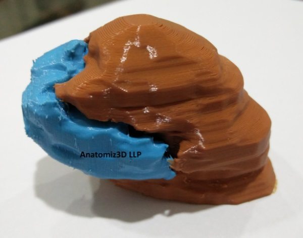
“In this case, an MRI was utilized for designing. The tongue and tumour were segmented as two different parts and printed in two different colours for easy identification. The 3D printed model helps understand the 3-dimensional anatomy of the part in question, and the depth, position and size of the tumor with respect to the tongue.”
By using 3D technologies, surgeons were better equipped to visualize the tumor and plan an effective and minimally invasive removal. Additionally, the 3D printed modeled proved helpful to the patient. Doctors were able to give a visual and physical representation of the procedure, which better prepared the patient for post-surgical rehabilitation processes.