Researchers from the Graz University of Technology (TU Graz) and the Medical University of Graz in Austria, have devised an automated system for cranial implant (or cranioplasty) design.
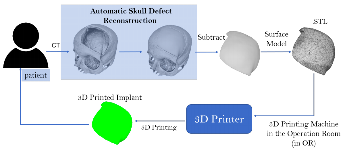
The program, which has been integrated into TU Graz’s Studierfenster cloud-based platform, uses deep learning algorithms to automatically restore the missing part of a skull. An STL file for a patient-specific implant can summarily be generated by subtracting the defective skull from the completed one. This process allows implants to be 3D printed on-site, and drastically lowers the existing lead times of producing them for routine surgeries, in addition to reducing the pain and discomfort of the patient.
3D printing cranial implants
Skull defects are often the result of head traumas, previous surgeries related to skull deformity correction or brain tumor removal, during which the surgeons need to remove a part of the cranial bone to access the brain. In order to repair this damage, a synthetic substitute is usually created using titanium or biocompatible polymers, because the original bone structure is too damaged or contaminated to be re-used.
The current production process used in the resulting cranioplasty, primarily relies on high-quality implant design and the manufacturing of cranial implants by professional companies, which can be expensive and time consuming. The high-level of expertise required to carry out the procedure, combined with the commercial software needed to optimize it, drive these costs even higher. Moreover, the human cost of the procedure also warrants consideration. Under current processes, the patient requires at least two operations: the craniotomy and the cranioplasty, which causes them additional suffering. This can take several days to take place, all of which the patient has an incomplete skull.
While advances in 3D bioprinting have accelerated the manufacturing of medical implant devices, there remains a need for a rapid, low-cost alternative. For instance, in a case study referenced by the researchers, a Spanish patient had to wait for an implant to be designed using several softwares, and be manufactured in the UK, before it was shipped for surgery. The research team used instances such as this to identify the existing implant design process as the primary bottleneck within the current clinical workflow.
The Graz University of Technology’s online platform
One method of reducing costs and lead times in the production process, would be to develop an ad hoc, free CAD software, for cranial implant design. This would still require a high-level of expertise to design and operate, so the researchers concluded that a low-cost fully-automatic on-site design and manufacturing software was the ideal solution. Moreover, automation of design remained a challenge. The 3D printed implant needed to fit precisely within the defected region of the skull, and provide the level of protection to the brain that its predecessor had done. If the implant’s boundary, thickness and shape consistency weren’t recreated perfectly, the part could leave the patient vulnerable to summary injuries.
The Studierfenster website was launched by two TU Graz students as a result. A cloud-based, open-science platform for medical image processing, the software can be accessed via any web browser. Numerous features have been added to the platform since its initial release, such as 3D face reconstruction from a 2D image, inpainting and restoration of aortic dissections, automatic aortic landmark detection and automatic cranial implant design. Most of the algorithms behind these interactive features run on the server, and they can be easily accessed using a common browser interface, allowing the platform to be used on smaller devices with lower computational capabilities.
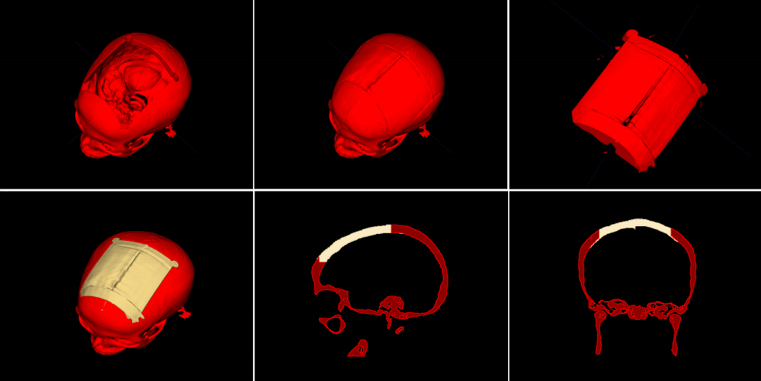
Studierfenster’s automatic cranial implant design system has been incorporated into the software since its early development phase, and was iteratively optimized from then onwards. The program works by uploading CT scans of the patient before and after the craniotomy into the software, which generates the implant by calculating the difference between the two skulls. This produces a surface model which is automatically 3D printed on-site in the hospital, removing the additional lead times and costs of external manufacturing. In addition, the program’s ability to subtract the defective skull from the reconstructed skull volume, allows the process to take place automatically, unlike prior efforts.
The research team concluded that their online system for automatic cranial implant design could substantially improve the current clinical routine around cranioplasty. There are drawbacks to the platform, and its effectiveness remains somewhat hardware and internet-dependent, while the conversion of the skull volume to a mesh can be slow. Nonetheless, the researchers remain confident that although the software is currently being used for educational and research purposes, it has the potential to be used in clinical settings in the future.
Cranial innovations in additive manufacturing
3D printing has been utilized to aid cranial reconstruction surgery consistently over the last few years. Researchers from Northwestern University, and the University of Illinois at Chicago (UIC) for instance, used 3D printed hyperelastic bone to regenerate skull defects in rats. The research could lead to the development of a cost-effective solution for craniofacial bone grafts.
Researchers from Texas A&M University combined 3D printing, biomaterial engineering and stem cell biology to create new, more efficient, customizable bone grafting materials. Using this technique, the scientists produced 3D printed highly-osteogenic scaffolds, that facilitate bone cell growth and serve as a sturdy platform for bone regeneration in custom shapes.
The UK’s North Manchester General Hospital (NMGH) used its 3D printing lab to create DCIA bone grafts for reconstructing the upper or lower jaws of cancer patients. The process yielded huge time and cost savings for the hospital.
The researchers’ findings are detailed in their paper titled “Hydrogel-Based Bioinks for 3D Bioprinting in Tissue Regeneration” published on June 1st 2020. The study was co-authored by Jianning Li, Antonio Pepe, Christina Gsaxner and Jan Egger.
You can now nominate for the 2020 3D Printing Industry Awards. Cast your vote to help decide this year’s winners.
To stay up to date with the latest 3D printing news, don’t forget to subscribe to the 3D Printing Industry newsletter or follow us on Twitter or liking our page on Facebook.
Looking for a job in the additive manufacturing industry? Visit 3D Printing Jobs for a selection of roles in the industry.
Featured image shows the Austrian researchers’ automated process, which allows users to design cranial implants. Image via the Graz University of Technology.
