3D bioprinting could be the exact tool that researchers need to discover the cause of Alzheimer’s disease and strokes.
Though animal models exist for the purpose of neurovascular research, a technical review written by a team at the University of Manchester in the UK makes the case for human cell models that can be studied at scale to pinpoint defective cells.
According to the paper, the quality of test models made to mimic the structure of the brain “increased significantly” when using bioinks and 3D bioprinting, an important discovery on the road to find a therapy and potential cure for debilitating and life-threatening conditions.
Identifying the cause of dysfunction
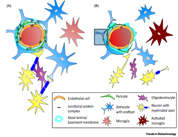
The University of Manchester’s lit review focuses on the replication of the neurovascular unit (NVU), a cluster of around 7 different cell types responsible for maintaining a healthy brain.
Typically, NVU cells combine to make the blood-brain barrier (BBB) that separates blood flow from brain fluid. In a defective brain, these cells have been identified as a potential contributor the neural dysfunction.
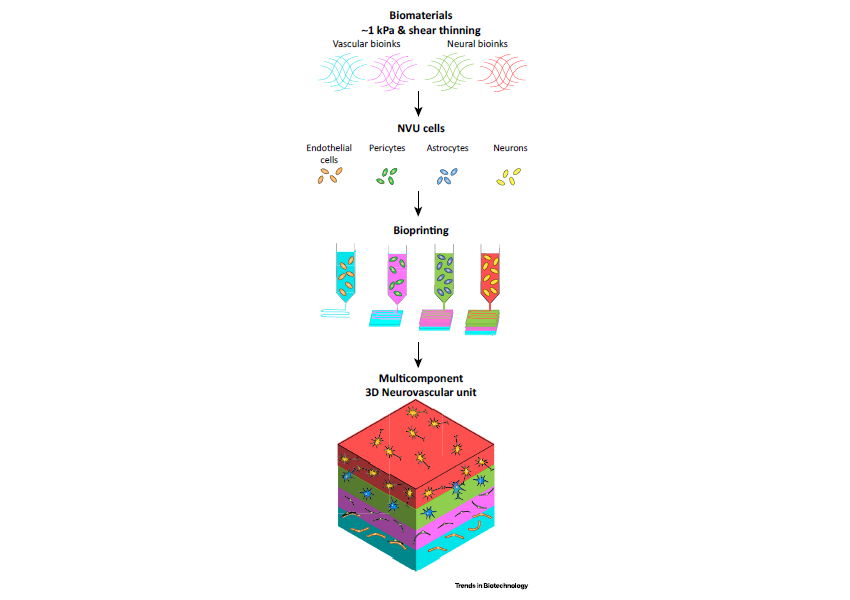
The layering of hydrogel mixtures, which are conducive to the culture of living cells, present scientists with a promising mode of faithfully recreating NVU structure.
In the review both direct and in-direct 3D bioprinting methods are assessed for their potential to mimic the NVU structure of the brain.

Indirectly 3D bioprinting the brain
Indirect 3D bioprinting is the process of casting a model from a 3D printed master mold. This method avoids any shear stresses associated with the 3D printing process therefore protecting sensitive, living material from harm.
It has been used variously to make microfluidic devices and prototype slow-release vaccinations. However by relying on pouring, materials cannot be deposited with any specificity.
Direct 3D bioprinting of the brain
Direct 3D bioprinting gives researchers the precision required to make an accurate medical model. It allows structures to have a further level of complexity, e.g. creating free standing scaffolds for cells, and aids complex multimaterial models. This is the method favoured by vascular 3D bioprinting at Harvard University.
According to the authors, “This approach is ideal for the manufacture of NVU models, where an optimal model requires both neural and vascular cell-laden ECM, with an interface for cell–cell interactions.”
“Bioprinting also enables the development of a reproducible model for comparative experiments between healthy and diseased cell types.”

A significant increase in brain quality
The University of Manchester’s medical review concludes explaining “Both direct and indirect printing techniques are able to create channels within the 3D construct […] enabling the fabrication of a BBB or vascular network.”
However, “By utilising bioinks and 3D bioprinting, the quality of the NVU model is increased significantly, which will lead to a better understanding of the molecular and cellular interactions between the different cell types in the NVU and how these go wrong in diseases associated with neurovascular dysfunction.”
The full review, ‘Tissue Engineering 3D Neurovascular Units: A Biomaterials and Bioprinting Perspective‘ is published online in Trends in Biotechnology journal. It is co-authored by Geoffrey Potjewydlink, Samuel Moxonlink, Tao Wang, Marco Domingos and Nigel M. Hooper.
Stay abreast of all the latest 3D printing research progress – subscribe to the 3D Printing Industry newsletter, follow us on Twitter, and like us on Facebook.
3D Printing Industry Jobs is live. Post a job or discover your next career move now.
Protolabs is sponsoring the 2018 3D Printing Industry Awards design competition. Submit your entries now with the chance of winning a 3D printer.
Featured image shows PET image of an adult male brain. Image by Jens Maus


