
Scientists working with the award winning Lewis Lab at Harvard University have made a significant advancement in the development of 3D bioprinted kidney models. Building on foundational work published in 2016, a new paper in the Proceedings of the National Academy of Sciences (PNAS) discusses the absorption of substances in 3D bioprinted models of a kidney’s promixal tubule.
Lab lead Professor Jennifer Lewis, who is also core faculty member of Harvard’s Wyss Institute for Biologically Inspired Engineering, comments, “Our new 3D kidney model is an exciting advance as it more fully recapitulates the proximal tubule segments found in native kidney tissue.”
“Beyond its immediate applications for drug screening and disease modelling, we are also exploring whether these living devices can be used to augment kidney dialysis.”
3D bioprinting for better drug screening
Proximal tubules are the microscopic segments of a kidney responsible for regulating the pH of filtrate. Part of the kidney’s nephron, the cell walls of proximal tubules can reabsorb valuable nutrients from the filtrate back into the kidney, and also redirect waste for secretion. By 3D printing these minute structures, the Lewis Lab’s aim is to recreate the behavior of kidney tissue for further study outside of the body. Eventually, this could lead to the blue-sky view of bio-engineered organs for transplant. More pressing though, is these models’ potential impact on pharmaceutical development.
The independent perfusion of a 3D bioprinted proximal tubule and vascular channel running in parallel. Clip via Wyss Institute at Harvard University
Annie Moisan, study co-author and Principal Scientist at the lab’s industry partner Roche Innovation Center Basel, explains, “Our system could enable the screening of focused drug libraries for renal toxicity and thus help reduce animal experiments.”
“I am thrilled by the continued efforts from us and others to increase the physiological relevance of such models, for example by incorporating patient-specific and diseased cells, since personalized efficacy and safety are the ultimate goals of predicting clinical responses to drugs.”
Simulating the activity of diabetes
Previously, the Lewis Lab had managed to 3D bioprint a singular, perfusable proximal tubule. The key development in this new paper is the addition of a perfusable blood vessel that runs alongside the tubule.
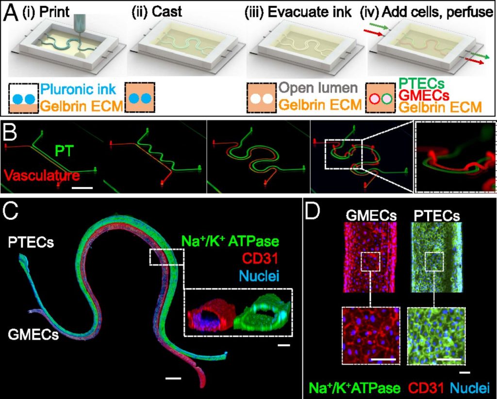
Both of the 3D bioprinted structures contain living cells. In experiments, the team studied the tubule’s ability to move glucose into the blood vessel. The team also simulated the occurrence of hyperglycemia – the high glucose levels that occur in diabetes patients. “We found that high levels of glucose transported to endothelial cells in the vascular compartment caused cell damage,” said study co-author and Lewis Lab Research Associate Kimberly Homan.
“By circulating a drug through the tubule that specifically inhibits a major glucose transporter in proximal tubule epithelial cells, we prevented those harmful changes from happening to the endothelial cells in the adjacent vessels.”
Further, as discussed in the body of the study, “Although these experiments focused on acute glucotoxicity, we believe that longitudinal studies carried out under conditions that mimic chronic hyperglycemia may provide valuable insights into treatments of diabetic vascular diseases.”
“Renal reabsorption in 3D vascularized proximal tubule models” is published open-access in PNAS journal. It is co-authored by , , , , , , and
For more of the latest 3D bioprinting research, subscribe to our 3D printing newsletter. You can also join us on Facebook and Twitter. Looking for a job in the industry? Then visit our 3D Printing Jobs board.
Featured image shows 3D bioprinting a proximal tubule (green) and blood vessel (red). Image via Wyss Institute at Harvard University
