bodyTop

3D printing to help diagnose patients with anemia

Access to healthcare and treatment and also diagnostic facilities can be difficult in remote regions. From rural areas to underdeveloped countries, not everyone lives in an area where a doctor or hospital can be reached in a reasonable amount of time. And delay in access to healthcare can lead to further complications when it comes to diagnosis and eventual treatment.

Development of a low cost test that combines smartphones and 3D printing is underway by a team of researchers from Kansas State University. The 3D printed auto-mixing chip will enable rapid smartphone diagnosis of anemia, and will potentially lead to broader uses, according to a new paper published by the team.
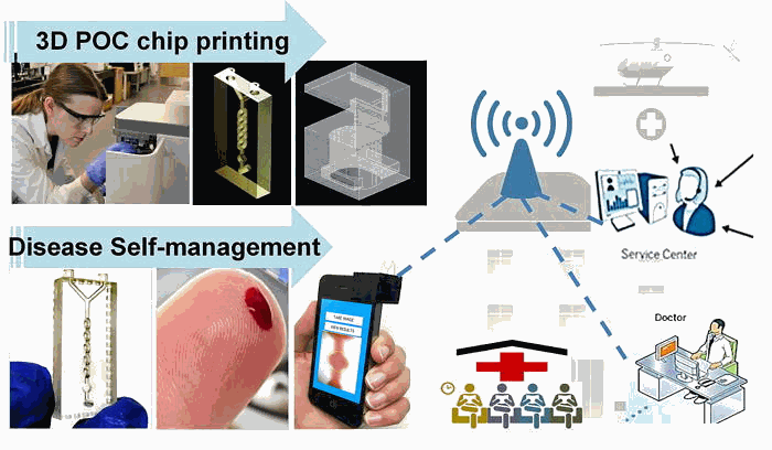
Low-cost, smartphone-based, 3D-printed POC microfluidic chip (smartphone iPOC3D system) for rapid diagnosis of anemia in 60 s.
Low-cost, smartphone-based, 3D-printed POC microfluidic chip (smartphone iPOC3D system) for rapid diagnosis of anemia in 60 s. Image via Biomicrofluidics/Kansas State University.

We first wrote about the team earlier this year, and since then they have published a paper in the journal of Biomicrofluidics. The journal article details exactly how their technology works, and how it will benefit the wider public it was made for.

The 3D printed auto-mixing chip developed integrates 3D design and 3D printing of microfluidic point-of-care (POC) device with smartphone-based disease diagnosis in one process as a stand-alone system. This could have further positive implications in the future, with the technology being adaptable for a number of other blood-based tests.
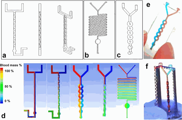
Design of three microfluidic mixers: (a) SAR, (b) serpentine channel, and (c) ring-shaped channel. Channel diameter was ∼500 μm. (d) 3D CFD simulation of blood mixing profile in three micromixers: Red flow is blood mixed with aqueous solution (blue); green color indicates complete mixing. (e) Experimental image of 3D printed planner micromixer showing incomplete mixing under capillary force using colored dye solutions. (f) Experimental image of 3D printed, 3D-structured micromixer showing efficient mixing within 1 s using colored dye solutions.
Design of three microfluidic mixers: (a) SAR, (b) serpentine channel, and (c) ring-shaped channel. Channel diameter was ∼500 μm. (d) 3D CFD simulation of blood mixing profile in three micromixers: Red flow is blood mixed with aqueous solution (blue); green color indicates complete mixing. (e) Experimental image of 3D printed planner micromixer showing incomplete mixing under capillary force using colored dye solutions. (f) Experimental image of 3D printed, 3D-structured micromixer showing efficient mixing within 1 s using colored dye solutions. Image via Biomicrofluidics/Kansas State University.

The device was modeled on the AutoCAD 360 app and printed on D3 ProJet 1200 using VisiJet®FTX Clear resin. It enables the detection of hemoglobin levels in blood, which helps identify iron deficiency that leads to anemia.

The test can be done in just 1 second, with the auto-mixing of reagents with blood via capillary eliminating the need for external pumps. Testing the device with a training set of patients consistently presented the same levels of diagnostic sensitivity and specificity as one would find with standard clinical machines.
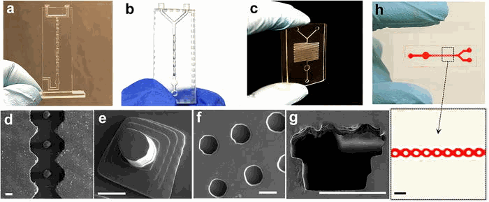
Characterization of 3D printing for fabricating microfluidic mixers: (a) SAR microfluidic mixer, (b) 3D ring-shaped micromixer, (c) and serpentine micromixer. Channel diameter is ∼500 μm. SEM imaging was used to characterize variable 3D-printed microstructures: (d) posts in curved microfluidic channel, (e) pyramid post arrays, (f) microwells, and (g) a D-shaped hollow microchannel. Scale bar: 200 μm. (h) The bonded layer of a 3D-printed open channel with a glass slide.
Characterization of 3D printing for fabricating microfluidic mixers: (a) SAR microfluidic mixer, (b) 3D ring-shaped micromixer, (c) and serpentine micromixer. Channel diameter is ∼500 μm. SEM imaging was used to characterize variable 3D-printed microstructures: (d) posts in curved microfluidic channel, (e) pyramid post arrays, (f) microwells, and (g) a D-shaped hollow microchannel. Scale bar: 200 μm. (h) The bonded layer of a 3D-printed open channel with a glass slide. Image via Biomicrofluidics/Kansas State University.

This device is incredibly low cost, with each test’s estimated cost being just 50 cents. Considering the components of this device other than a smartphone are 3D printed, it can be easily adopted and help bring mobile diagnostic services to where they are needed most.