One of the biggest hurdles to the 3D printing of complex human tissue, complete organs in particular, is the ability to 3D print blood vessels capable of carrying the necessary blood supply to bioprinted cells. Just as various businesses and research institutions have demonstrated the ability to fabricate more and more types of human tissues, however, we have seen an increasing number produce 3D printed blood vessels. Last week, Revotek, in China, unveiled a machine designed specifically for such a purpose. And, today, Rice News reports that new research has been published by Rice University and the University of Pennsylvania demonstrating another method for delivering blood to artificial tissues.
In an article published in the journal Tissue Engineering Part C: Methods, the team, led by assistant professor of bioengineering at Rice Jordan Miller and assistant professor of surgery at Penn Pavan Atluri, were able to create a silicon construct with a complex network of blood vessels, in which blood was able to flow normally to surgically attached, native blood vessels. While tissue engineers have, in the past, implanted engineered tissue and waited for the body to grow its own blood vessels to supply oxygen and nutrients to the tissue, Miller’s team instead opted fabricating the blood vessel network itself. As a result, there’s no need to wait for a native network to form, reducing the possibility that cells within the tissue die from a lack of oxygen.
Miller tells Rice News, “We had a theory that maybe we shouldn’t be waiting. We wondered if there were a way to implant a 3D printed construct where we could connect host arteries directly to the construct and get perfusion immediately. In this study, we are taking the first step toward applying an analogy from transplant surgery to 3D printed constructs we make in the lab.” He adds, “What a surgeon needs in order to do transplant surgery isn’t just a mass of cells; the surgeon needs a vessel inlet and an outlet that can be directly connected to arteries and veins.”
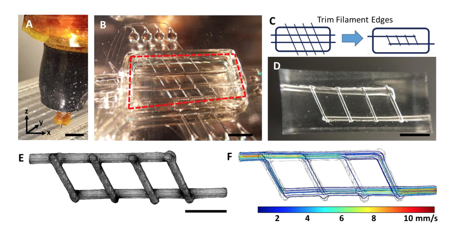
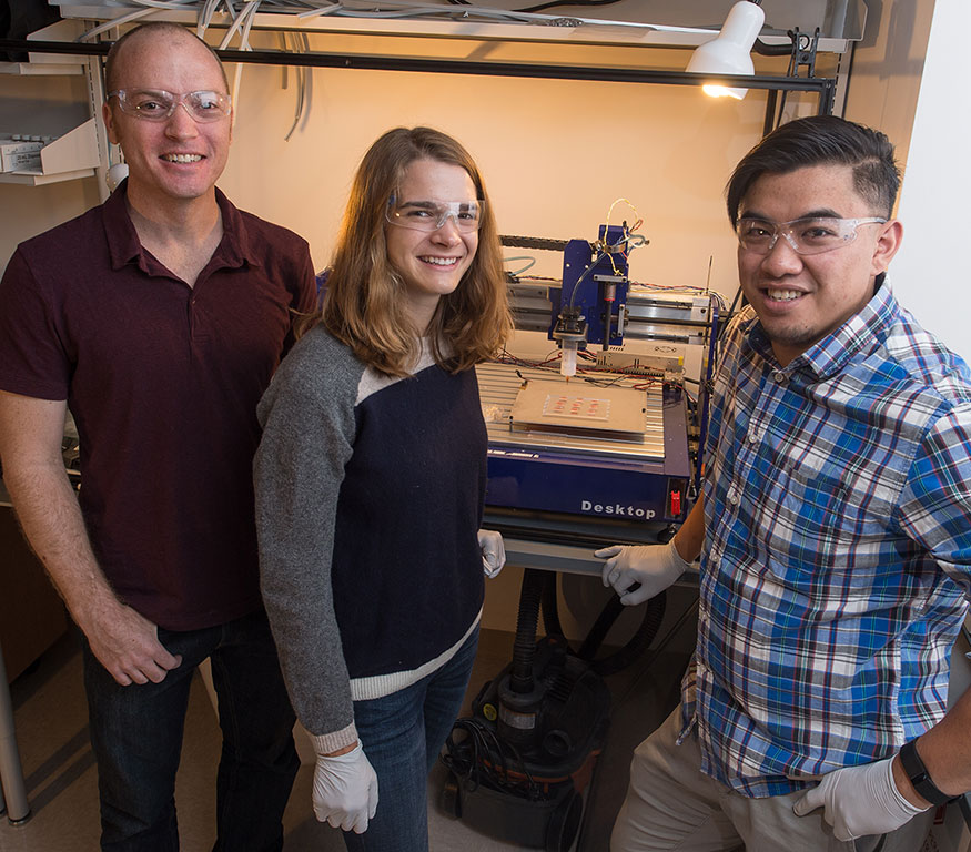
To create the vessel network, Bioengineering graduate student Samantha Paulsen and research technician Anderson Ta first 3D printed a positive for the silicone casting of the vessel network, according to Rice News. 3D printing the positive in sugar glass with an open source printer, the team then put their hardened sugar print into a mold that was later filled with silicone gel. In turn, the sugar was dissolved, while the silicone blood vessel network remained in tact.
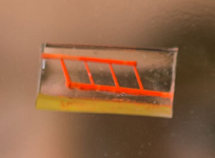
Miller continues, “They don’t yet look like the blood vessels found in organs, but they have some of the key features relevant for a transplant surgeon. We created a construct that has one inlet and one outlet, which are about 1 millimeter in diameter, and these main vessels branch into multiple smaller vessels, which are about 600 to 800 microns.”
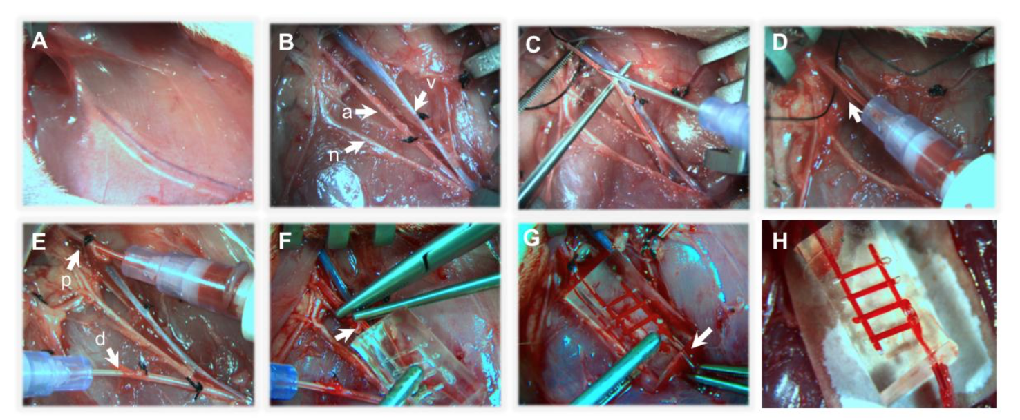
Once the proof-of-concept was achieved, the Rice team worked with Atluri’s surgeons to surgically implant the vessel network into a lab rat, used as an animal model, where blood flowed without issue for up to three hours. The study was deemed a complete success, with Miller saying, “This study provides a first step toward developing a transplant model for tissue engineering where the surgeon can directly connect arteries to an engineered tissue. In the future we aim to utilize a biodegradable material that also contains live cells next to these perfusable vessels for direct transplantation and monitoring long term.” Specifically, the team will use endothelial cells to reduce clotting and improve the transportation of nutrients in the blood vessels, as well as improve the transportation of nutrients. They may also try using different hydrogels for the fabrication of the blood vessels in the future.
Now that the team has demonstrated the successful implantation of an artificial blood vessel network, they can work to bring blood flow to bioficial tissues. With the ability to pump oxygen and nutrients to these tissues, they may even be able to expand the number of cell types that can be viably supported in the lab, slowly growing their research until they’re able to construct complete bioprinted organs. At that point, bioprinting will no longer be a tool for research, but for saving lives.