
Researchers from the University of Wisconsin-Madison (UW-Madison) have developed a novel approach for 3D printing functional human brain tissue.
The 3D printing process can create active neural networks in and between tissues that grow in a matter of weeks.
The researchers believe that their 3D bioprinted brain tissue provides an effective tool for modeling brain network activity under physiological and pathological conditions, and can also serve as a platform for drug testing.
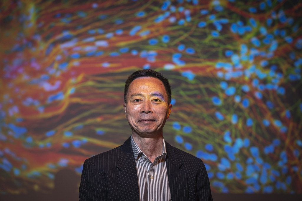
“This could be a hugely powerful model to help us understand how brain cells and parts of the brain communicate in humans,” explained study co-author Su-Chun Zhang, professor of neuroscience and neurology at UW–Madison’s Waisman Center. “It could change the way we look at stem cell biology, neuroscience and the pathogenesis of many neurological and psychiatric disorders.”
The study, titled ‘3D bioprinting of human neural tissues with functional connectivity,’ was published in the latest edition of the journal Cell Stem Cell.
3D bioprinting functional brain tissue
According to the scientists, current methods for creating functional human brain tissue are limited. Stiff scaffolds are commonly 3D printed to support the complex structure, which the brain cells are then seeded into to grow the human tissue.
This process usually results in neural cells being unevenly distributed, instead forming thick cell clusters. Moreover, as 3D printed scaffolds are usually not biodegradable, they block cell migration, preventing active neural networks from forming between layers.
The new approach overcomes these challenges by 3D printing the brain tissue horizontally, rather than the conventional vertical extrusion approach. To prevent the 3D printed cell bands from mixing, thrombin was added as a crosslinking agent after the tissue was deposited.
The scientists also employed a softer bio-ink than previous approaches, allowing the cells to form connections both inside and between tissue layers, more comparable to a real human brain.
“The tissue still has enough structure to hold together but it is soft enough to allow the neurons to grow into each other and start talking to each other,” said Zhang. “Our tissue stays relatively thin and this makes it easy for the neurons to get enough oxygen and enough nutrients from the growth media.”
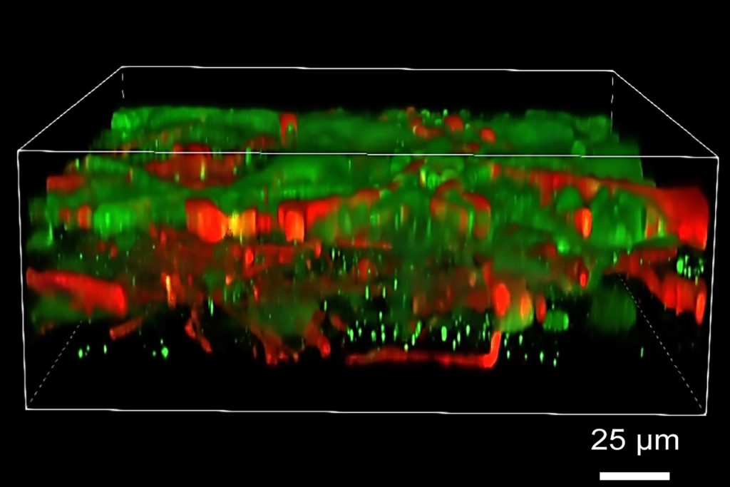
Once 3D printed, the neurons could communicate, send signals, and interact with each other through neurotransmitters. The novel structures even created proper networks with support cells that were added to the 3D printed tissue
“We printed the cerebral cortex and the striatum and what we found was quite striking,” added Zhang. “Even when we printed different cells belonging to different parts of the brain, they were still able to talk to each other in a very special and specific way.”
The UW-Madison team used a commercially available 3D bioprinter during the study, which Zhang claims could be further modified to 3D print specific types of brain tissue on-demand in the future.
The team also states that its new 3D printing technique is accessible to other research labs, given that it does not require special bio-printing equipment or culturing methods to keep the tissue healthy. The 3D printed tissue can also be easily studied in depth with commonly available microscopes, electrodes, and standard imaging techniques.
Ultimately, it is hoped that the 3D printed brain tissue could be used to study cell-cell signaling in Down syndrome, assess how healthy tissue interacts with Alzheimer disease-affect tissue, test new drug candidates, or even observe how the brain grows.
Zhang believes that the 3D printed tissue can be used to study “almost every major aspect of what many people at the Waisman Center are working on.”
“It can be used to look at the molecular mechanisms underlying brain development, human development, developmental disabilities, neurodegenerative disorders, and more,” added Zhang.
3D printing advances neurological research
The use of additive manufacturing and 3D bioprinting continues to play a key role in advancing medical research, treatment, and drug discovery. When surveyed on the future of 3D printing, 3D printing experts argued that the industry will continue see significant advancements in medical applications.
The UW-Madison team is not the first to explore the potential offered by 3D printing in the study of neurological conditions and the development of new treatments. Back in 2022, Researchers from the Federal University of Sao Paulo (UNIFESP) in Brazil developed a method for 3D printing brain cells that can survive for at least 14 days after fabrication.
The study, which was funded by a research grant from the Sao Paulo Research Foundation (FAPESP), resulted in a model that was more realistics to natural brain tissue than other protocols at the time.
The 3D printed tissue could be used to understand the functionality of neural cells in relation to central nervous system diseases. Additionally, the researchers leveraged their proprietary method to explore materials that could be used to repair brain areas damaged by traumatic injuries or strokes.
Elsewhere, Scientists from medical tech company Fluicell partnered with clinical R&D firm Cellectricon and the Swedish Karolinska Institutet university to 3D bioprint neural cells into complex patterns.
The team leveraged microfluidic 3D printheads featured on Fluicell’s Biopixlar 3D bioprinter to accurately arrange rat brain cells within 3D structures, without damaging their viability. The resulting 3D printed cerebral tissue could then be used to model the progress of neurological disease or test the efficacy of related drugs.
Subscribe to the 3D Printing Industry newsletter to keep up to date with the latest 3D printing news. You can also follow us on Twitter, like our Facebook page, and subscribe to the 3D Printing Industry Youtube channel to access more exclusive content.
Are you interested in working in the additive manufacturing industry? Visit 3D Printing Jobs to view a selection of available roles and kickstart your career.
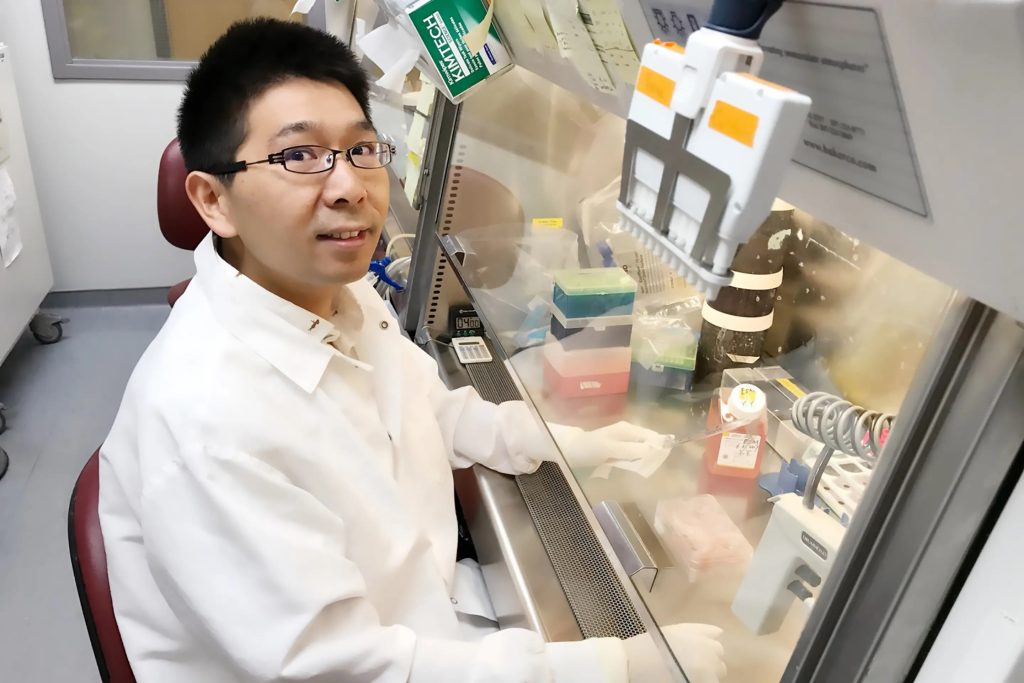
Featured image shows the study’s lead author, Yuanwei Yan, in the Zhang lab at UW–Madison. Photo via UW-Madison.