Researchers from Northwestern University and the Ann & Robert H. Lurie Children’s Hospital of Chicago have published a paper detailing the ongoing development of ink for 3D printing bioprosthetic ovaries.
This ink is imbued with structural proteins derived from a pig ovary. The location of these proteins has been mapped and identified in the study.
Using the ink, it may be possible to 3D print artificial ovaries that can be implanted in infertile women, subsequently allowing them to bear children. “This is a huge step forward for girls who undergo fertility-damaging cancer treatments,” comments senior author Monica Laronda, Ph.D., Director of Basic and Translational Research, Fertility & Hormone Preservation & Restoration Program at Ann & Robert H. Lurie Children’s Hospital of Chicago, and Assistant Professor of Pediatrics at Northwestern University Feinberg School of Medicine.
“Our goal is to use the ovarian structural proteins to engineer a biological scaffold capable of supporting a bank of potential eggs and hormone producing cells. Once implanted, the artificial ovary would respond to natural cues for ovulation, enabling pregnancy.”
3D printing functional ovaries
Ann & Robert H. Lurie Children’s Hospital is a nationally ranked pediatric specialty hospital based in Chicago. The hospital’s research output is conducted through the Stanley Manne Children’s Research Institute, which is focused on “improving child health, transforming pediatric medicine and ensuring healthier futures.” The Ann & Robert H. Lurie Children’s Hospital is also the training ground for Northwestern University Feinberg School of Medicine.
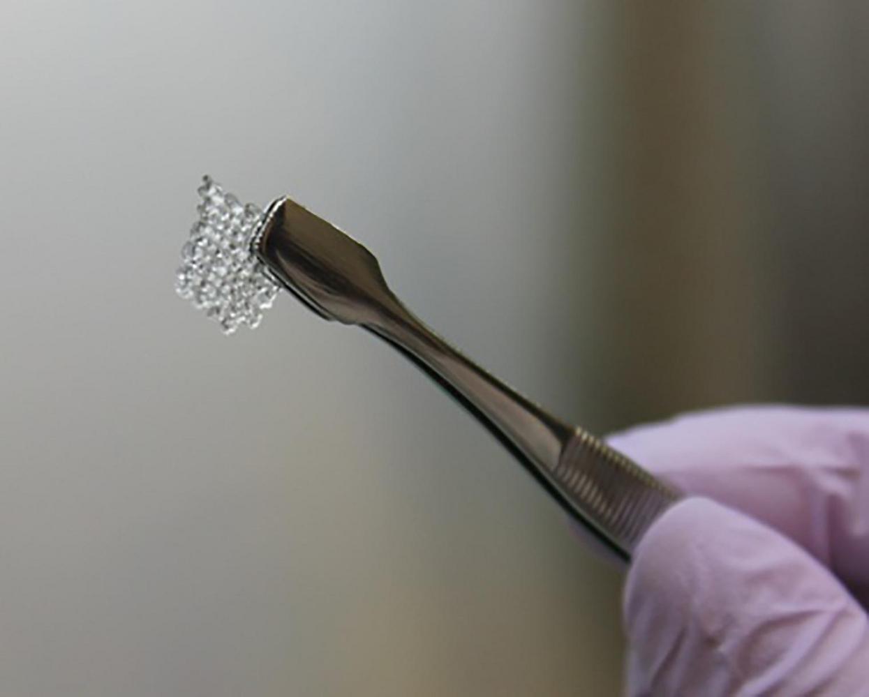
In 2016, Dr. Laronda, one of the authors of the new study, revealed details surrounding the work of her research team in 3D printing and successfully implanting, a functional bioprosthetic ovary in a female mouse. This was done using 3D bioprinted scaffolding populated with ovarian follicles, created from a biogel that was derived from animal protein collagen.
In 2017, details of the research were published in a paper in Nature Communications. The mouse was able to ovulate and give birth to a healthy litter with a 3D printed organ, which replaced the mouse’s original ovaries. The main aim of the research project was to demonstrate the possibility of restoring fertility and hormone production in women unable to do so following cancer or other development issues.
Towards a bio-ink for 3D printing human ovaries
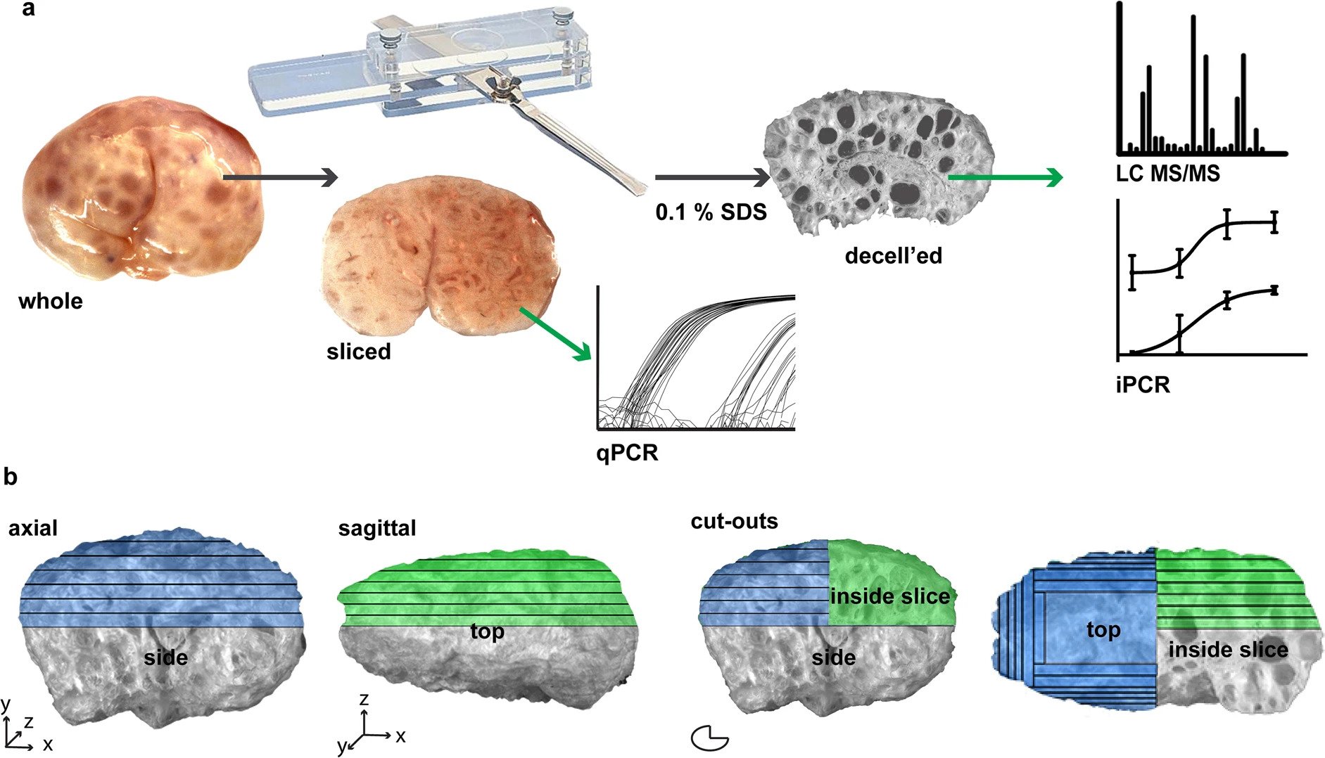
Dr. Laronda and three other colleagues received a patent for the creation of an artificial ovary in November 2019. In the new study, the researchers map out the composition of pig ovaries, providing a pipeline for identifying the location of its structural proteins.
“The structural proteins from a pig ovary are the same type of proteins found in humans, giving us an abundant source for a more complex bio-ink for 3-D printing an ovary for human use,” adds Dr. Laronda. “We are one step closer to restoring fertility and hormone production in young women who survive childhood cancer but enter early menopause as a late effect. There are still several steps to go and we are excited to test our new inks.”
The research paper also explains how the methodology used to map the structural proteins can be applied by scientists who are investigating other human organs as well. Northwestern University continues to maintain a strong output in research relating to 3D printing technology. The university recently detailed a new large scale SLA 3D printer that can print half a yard (457.2 mm) in an hour, a reportedly record-breaking throughput in 3D printing. Additionally, researchers from the academic institution have also used 3D printed hyperelastic bone to regenerate skull defects in rats, which can potentially lead to the development of a cost-effective solution for craniofacial bone grafts.
The study, titled ‘Proteomic analyses of decellularized porcine ovaries identified new matrisome proteins and spatial differences across and within ovarian compartments,’ is published in Scientific Reports. It is co-authored by Nathaniel F. Henning, Richard D. LeDuc, Kelly A. Even, and Monica M. Laronda.
Subscribe to the 3D Printing Industry newsletter for the latest news in additive manufacturing. You can also stay connected by following us on Twitter and liking us on Facebook.
Looking for a career in additive manufacturing? Visit 3D Printing Jobs for a selection of roles in the industry.
Featured image shows scientist holding a 3D printed mouse ovary cell scaffold. Photo via Northwestern University.