
Researchers at the University of St Andrews in Scotland have discovered two new techniques for creating 3D images of internal, biological tissues.
The non-invasive 3D scanning methods go deeper into tissue than ever before, and are capable of collecting a complete 3D image without rotating or taking multiple images of a specimen.
The technology has many implications for the field of diagnostics, and is hoped to help advance the understanding of complex conditions including cancer, Alzheimer’s disease and Parkinson’s.
The future of medical imaging?
Medical is one of the largest markets for innovations 3D scanning and 3D printing. Anatomical models made using CT and MRI scan data are is one of the most common applications, and are proving lucrative services for companies such as Stratasys and 3D Systems.
The way 3D medical data is handled is also of paramount importance. Materialise, in collaboration with Siemens Healthineers, is leveraging its Mimics inPrint software to bring 3D printing capabilities to more radiology departments around the world.
The Materialise Mimics Innovation Suite has also become the first software to receive FDA clearance for intended use in 3D printing anatomical models for diagnosis, and research, including this report from researchers in Jiangsu Sheng, China, proposes a future where 3D scanners replace CTI and MRI scans.
This St. Andrews study could be an important step towards future hardware that tailors to burgeoning medical innovation. If it can be used in a place of an MRI or CT scan, it could make the diagnosis a lot more comfortable for the patient.
Getting a clearer image
Dr. Jonathan Nylk is a researcher at St. Andrews School of Physics and Astronomy who worked on the new 3D imaging methods. Explaining the techniques, Dr. Nylk says, “We’ve recently discovered particular beam shapes that retain their shape when travelling through biological tissue.”
“These beams, called Airy beams and Bessel beams resist the effects of scattering.”
Airy and Bessel beams are commonly used in imaging techniques. The breakthrough in the St. Andrews research is that the team have developed techniques that mean the beams can retrieve image data from deeper within tissue.
As outlined by Dr. Nylk, though the beams resist the effect of scattering “they still become dimmer as they travel deeper, so it remains challenging to collect enough signal back through the tissue to form an image.”
Tissues slice by slice
The beam study improves upon the existing method of light-sheet microscopy.
Light-sheet microscopy is a high-speed imaging technique that delivers intermediate-to-high optical resolution. A microscope is used to direct razor-thin sheets of light on an object, and the resulting feedback delivers an image in slices. Adding Airy or Bessel beams to light-sheet microscopy has improved the depth of this method.
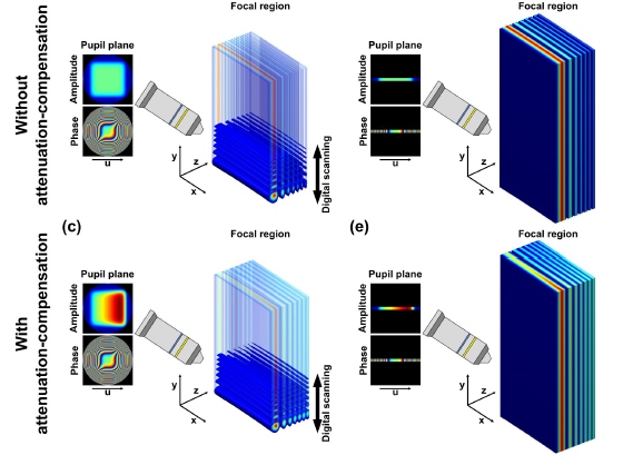
Dr. Nylk adds, “Now we show that these beams can be further enhanced to give us more control over their shape, such that they actually get brighter as they travel (propagate),”
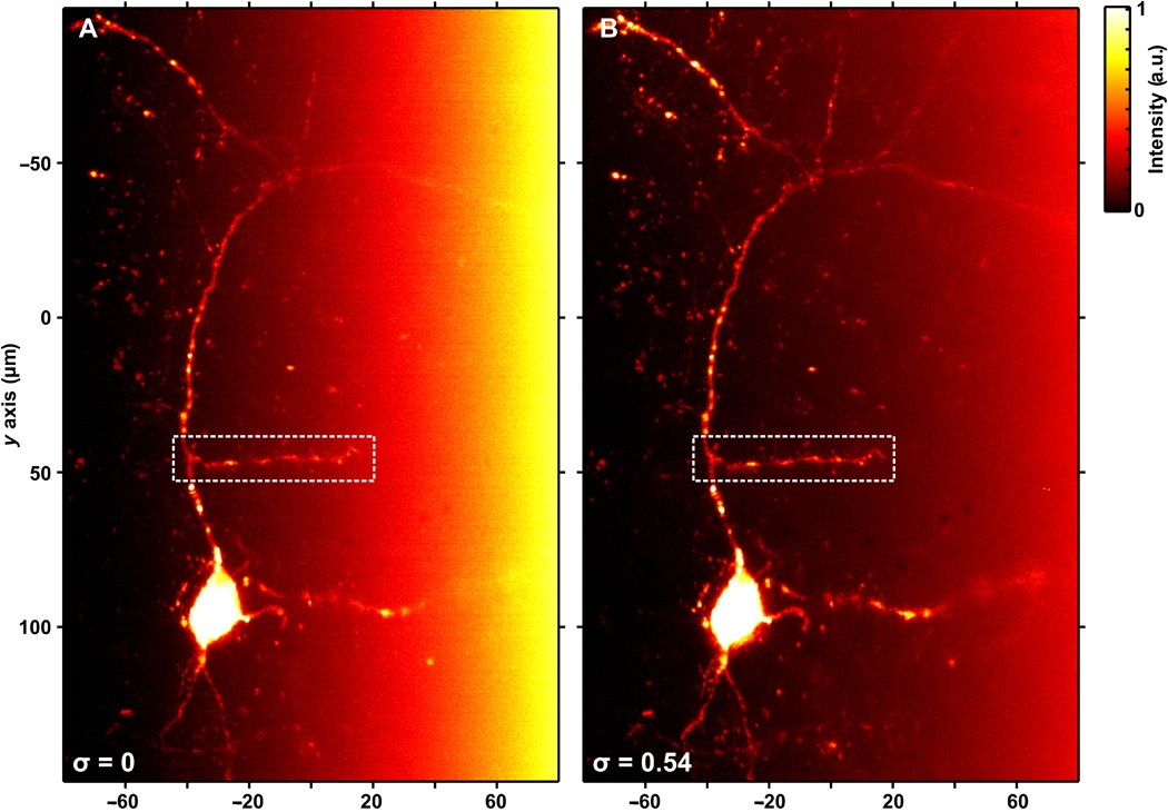
“When the increase in brightness (intensity) is matched with the decrease in brightness (intensity) when travelling through tissue, a strong signal and a clear image can still be acquired from deep within the sample.”
Airy light-sheets in particular were capable of delivering “sharp images over a volume ten times larger than previously possible.”
In addition, the Airy and Bessel beam breakthroughs could have implications for other 3D imaging techniques – maybe we’ll see the technology in a handheld scanner soon?
Light-sheet microscopy with attenuation-compensated propagation-invariant beams is publcihed online in Science Advances journal. It is co-authored by Jonathan Nylk, Kaley McCluskey, Miguel A. Preciado, Michael Mazilu, Zhengyi Yang, Frank J. Gunn-Moore, Sanya Aggarwal, Javier A. Tello, David E. K. Ferrier and Kishan Dholakia.
Stay up to date with all the latest research – subscribe to the 3D Printing Industry newsletter, follow us on Twitter, and like us on Facebook.
Vote now for 3D scanning application of the year and more in the 2018 3D Printing Industry Awards.
Find your next opportunity at 3D printing jobs and post a vacancy for free throughout April.
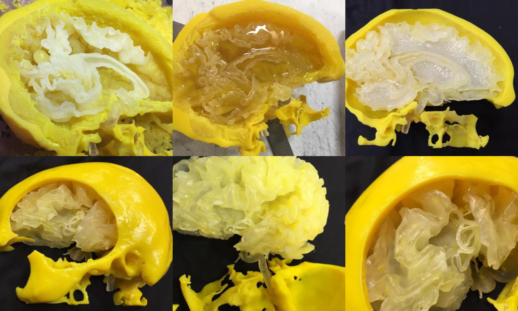
Featured image shows maximum intensity projections of Airy Light-Sheet Microscopy images. Image via Science Advances
