
A team of researchers from Carnegie Mellon University have developed a new method of 3D bioprinting that enables the production of realistic fully-sized human heart models.
The scientists’ Freeform Reversible Embedding of Suspended Hydrogels (FRESH) technique, involves extruding the eco-friendly alginate polymer into a custom-made gelatin container. Leveraging their novel process, the team aims to work with surgeons to create patient-specific clinical models for surgical training and pre-planning applications.
“The surgeon can manipulate it and have it actually respond like real tissue,” said Professor Adam Feinberg, who led the project. “So when they get into the operating site they’ve got an additional layer of realistic practice in that setting.”
“We can now build a model that not only allows for visual planning but allows for physical practice.”
A ‘FRESH’ new 3D printing approach
An increasing number of surgeons are adopting 3D printing as a means of developing bespoke models that allow them to explain cardiac procedures to their patients. Using bioprinting to produce these replicas enables them to be realistic, but also opens the possibility of tissue engineering and regenerative medicine applications in future.
At present, common 3D printing techniques such as stereolithography (SLA) and Fused Deposition Modeling (FDM) are being used to reproduce realistic-looking organs. Although such methods have generally yielded positive results, their wider adoption has so far been limited by their cost, and the level of expertise required to manufacture them.
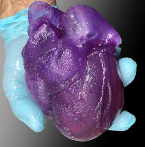
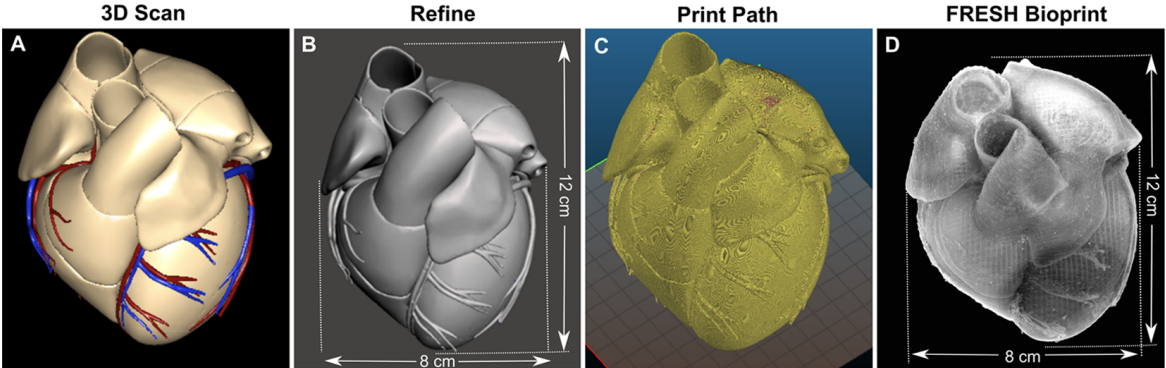
In order to overcome these limitations, Professor Feinberg and his colleagues have spent two years researching how to print a human heart model in full-size and came up with a FRESH new approach. The team’s novel technique begins with using data gathered from MRI scans and other scanning processes to design an accurate 3D model.
The resulting designs are then printed using a needle measuring 250 microns in diameter, which extrudes alginate into a custom-made container that’s large enough to fit a fully-sized replica. The team’s new method was ultimately found to yield more durable models than before, potentially allowing them to be used more effectively as a surgical training tool.
3D printing the fully-sized surgical models
During the 3D printing process, the scientists adopted alginate as the main material for their models because it closely resembles the texture and mechanical properties of organic cardiac tissue. What’s more, softer materials such as TPUs and silicone show deformations under the force of gravity, making the slightly more rigid alginate a better-suited alternative.
After fabricating a series of prototypes, the researchers post-processed them for 12 hours within a gelatin container, before placing them into an incubator overnight to remove their gelatin supports. Once the additive cardiac models were ready, Feinberg and his team proceeded to test them by seeing how far the polymer could be stretched while being stitched.
Results indicated that alginate featured sufficient tensile strength to be made into heart models for surgeons to practice on. The researchers then proceeded to use their FRESH approach to fabricate smaller models consisting of coronary arteries filled with fake blood that could be useful for training surgeons too.
Interestingly, the team revealed that the needle tip of the extruder was problematic, as it needed to be long enough to reach the bottom of the bath, as well as to support the printed material. To solve this issue, a 3D printed needle support collar was created which could be replaced and attached to the printer’s extruder head at will.
The researchers concluded that their 3D model was suitable as a surgical training tool for practising suturing, as well as other operations that could be conducted on a real human heart. Overall, the project could open up other avenues of research, and in future, the team hope that their FRESH approach leads to the development of a biomedicine testing tool.
3D printing and previous matters of the heart
In recent years, various scientists have adopted 3D printing as a means of producing realistic models of organs that broaden our understanding of the human body.
In July 2020, the University of Minnesota developed a new bio-ink that helped them create a functional 3D printed beating heart made out of cell-laden biomaterial. The team’s novel material enabled them to 3D print an aortic replica with more chambers, ventricles and a high cell wall thickness.
Earlier this year, The University of Texas at El Paso (UTEP) and biomedical researchers from Texas Tech University Health Sciences Center El Paso (TTUHSC El Paso) worked together to 3D print mini-hearts. The cell-laden structures were sent to the International Space Station (ISS) to research how microgravity affects the function of the human heart.
Similarly in 2018, a group of doctors from the Zhengzhou University carried out a study on a group of 25 patients to highlight how 3D printed anatomical models could be used in heart operations. The team eventually used CT scans to develop a patient-specific model that could be 3D printed at a 1:1 scale.
The research paper that includes the world’s first full-size 3D printed heart is titled FRESH 3D Bioprinting a Full-Size Model of the Human Heart. It is co-authored by Eman Mirdamadi, Joshua W. Tashman, Daniel J. Shiwarski, Rachelle N. Palchesko, and Adam W. Feinberg.*
Check us out on Twitter and Facebook for more updates! Don’t forget to subscribe to the 3D Printing Industry newsletter to keep up-to-date with the latest 3D printing news.
Are you looking for a job in the additive manufacturing industry? Visit 3D Printing Jobs for a selection of roles in the additive manufacturing industry.
Feature image shows the full-size 3D printed adult heart-model Feinberg and his team have manufactured. Photo via Carnegie Mellon University.