
Researchers from the UK-based Teeside University, have used 3D printing and scanning techniques to advance a key aspect of forensic investigation: Physical Fit Analysis (PFA). Utilizing and comparing two different 3D imaging methods, the research team were able to recreate human bone fragments for use within the PFA process. Not only could this prevent unnecessary damage from occurring to genuine evidence during a Crime Scene Investigation (CSI), but it may also expand on the forensic applications of 3D printing.
“Fused Filament Fabrication (FFF) 3D printing proved to be an accurate and useful method for creating physical replicas of the bone fragments, to perform physical fit analysis (PFA) and bone fragment reconstruction. We therefore recommend μCT imaging paired with FFF 3D printing, as an excellent option for non-destructive physical fit confirmation, when working with small fragments and burned bone,” said the research team.
Physical fit analysis in Crime Scene Investigation
CSI often requires investigators to examine a range of items as evidence, including human remains, some of which may be damaged or fragmented as a result of the trauma suffered during the event. These remains routinely go through PFA to determine whether they fit together. If this process leads to a positive physical fit, it could place suspects at the scene of a crime, or facilitate object reconstruction that potentially solves the case. Nonetheless, PFA involves a great deal of matching and manual handling, which can lead to the fragments becoming damaged during the process.
Moreover, there are instances where PFA can be extremely challenging, such as when fragments may pose a biological hazard, be extremely small, or the bone itself may be too fragile to move. Considering that the reconstruction process usually involves gluing the parts back together, this can cause problems, and prevent investigators from fully understanding the nature of the trauma. This makes certain-shaped bones difficult to document or present, particularly with those fragments that are three dimensional and complex in nature, or embedded in an external material. As a result, two-dimensional representations of such physical fit results are not always sufficient for presentation in courts or interpretation by experts.
While 3D scanning and modelling have been used in a range of forensic anthropology applications, the handling and reconstructing of bone fragments remains an issue. For instance, 3D modelling is currently used in dismemberment, weapon matching, craniometrics and facial reconstruction cases. Volume scanning has allowed for high resolution images to be obtained, on the nanometre scale in some instances, but they tend to be expensive, time consuming and require specialist expertise and software to operate. Surface scanning methods, on the other hand, tend to be cheaper, and more user-friendly, and are often used for postmortem quantitative injury analysis, landmarking, and the analysis of soft tissue injuries.
Additive manufacturing meanwhile, has proved accurate enough to produce dental models for aiding in maxillofacial surgery. Studies have also demonstrated the applicability of 3D printing for the visualisation and analysis of forensic evidence, and the researchers set about combining the technologies to create 3D printed skull fracture replicas. These 3D models offer the potential for PFA to take place without having to excessively handle the original evidential fragments, while minimising any damage or contamination risks. In addition, such models offer 360 degree visualisation in an engaging and understandable format, that could be used to improve jury comprehension during trials.
3D printing skull fragment replicas
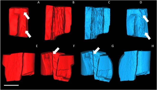
The researchers compared Micro Computed Tomography (μCT), a volume scanning technique, with structured light scanning (SLS) a surface scanning technique, to assess the pros and cons between the two methodologies. In order to test their potential for conducting PFA, the team 3D printed two models of burnt bone fragments, to simulate the damage that might be encountered in a genuine investigation.
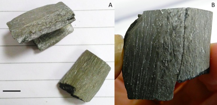
Modelled on an archaeological human femur donated by the University of Portsmouth, the replicated bone samples were cut and burned in a Gallenkamp Muffle Furnace at 600°C for 30−60 minutes. Each section of bone fragmented longitudinally, naturally, into at least two separate pieces, either during the burning process or during cooling. The two adjoining fragments were 3D imaged and printed to evaluate the techniques for their use in visualising and analysing the physical fit of burned bone fragments.
The SLS scanner used for testing was a Shining 3D EinScan Pro+, while the larger μCT ZEISS Xradia 520 Versa scanner was also chosen, due to its simple setup and non-destructive process. After scanning, the fragments were 3D printed with a FFF Prusa i3 desktop printer, using PLA filament. The optimal print quality (0.15 mm) was selected, and infill levels were set at 0% to create a completely hollow print. PFA was then conducted on the pairs of 3D printed bone models, with accuracy determined on the basis of feature matching and alignment between the two fragments, as well as the haptic ‘feel’ of the fit.
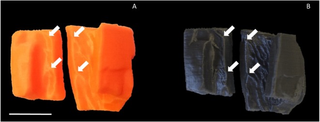
3D printing was found to preserve a high level of detail for both the μCT and SLS models, and overall, the prints produced were of a sufficient quality to perform PFA. Based on the ‘fit quality’ criteria set out by the team, the confirmation of physical fit was found to be easier using the μCT prints compared with the SLS prints. Moreover, in all fragment pairs created, the μCT models offered a closer and more robust fit, producing surface structures in greater detail, which was found to be of value in feature matching.
As a result, the researchers concluded that FFF 3D printing could be utilized to produce bone fragment replicas to a sufficient level of detail that either 3D scanning technique could be used. In addition, the team suggested that Selective Laser Sintering (SLS) 3D printing could prove to be a more efficient method of producing the models in future research, by negating the need for support structures used during FFF production. While it could ultimately prove more costly to implement, SLS printing would also result in a higher surface finish, according to the team. Additionally, the successfully reproduced bone fragments could open new applications for 3D printing in other aspects of the PFA process.
“The application of 3D imaging and printing for PFA has many advantages compared with traditional methods. Virtual reconstruction of highly fragmented, fragile, and potentially embedded remains, offers an opportunity to generate full reconstructions without compromising the original bone fragments,” concluded the researchers.
“Furthermore, 3D prints from particularly small fragments or bones with micro-scale details can be isometrically scaled up, generating 3D replicas to visualise fit and perform PFA on items that previously would have been extremely challenging.”
Additive advances in CSI
3D scanning and printing technology have been used in a range of ways to assist in CSI applications in recent years.
The Abu Dhabi Police Agency for example, have launched an additive manufacturing initiative to help solve crimes. Using 3D printing, the police force could produce dioramas that enable them to thoroughly assess a crime scene, or be summarily used to communicate a series of events in a court setting.
Police officers in Cascade County, Montana have started using FARO Focus 3D Laser Scanners to survey crime scenes. Capable of taking a full sweep of the scene, the 3D scanner potentially cuts down the personnel needed to record images by up to 80%.
Similarly, the UK-based West Yorkshire Police (WYP) force has installed a DeltaWASP 40 70 Industrial 3D printer at its Wakefield HQ. The machine is also used by its Regional Scientific Support Unit (SSU) in the recreation of crime scenes.
The researchers’ findings are detailed in their paper titled “Reconstruction and physical fit analysis of fragmented skeletal remains using 3D imaging and printing” published in the Forensic Science International journal. The report was co-authored by Amber J.Collings and Katherine Brown.
You can now nominate for the 2020 3D Printing Industry Awards. Cast your vote to help decide this year’s winners.
To stay up to date with the latest 3D printing news, don’t forget to subscribe to the 3D Printing Industry newsletter or follow us on Twitter or liking our page on Facebook.
Looking for a job in the additive manufacturing industry? Visit 3D Printing Jobs for a selection of roles in the industry.
Featured image shows the skull replicas that the research team produced using the μCT scanning method. Image via Science Direct.
