Prevention, as they say, is cheaper and easier than the cure. As such, 3D printign and scanning technologies are finding increasing application in medicine around the world for point-of-care diagnosis of various infections and conditions such as the zika virus and degenerative eye disease.
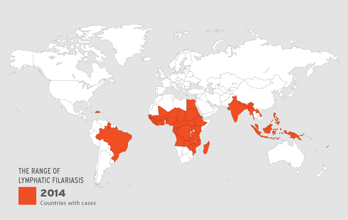
Using a 3D scanner developed by Atlanta startup LymphaTech, researchers in Sri Lanka and at Washington University School of Medicine in St. Louis have proved that the device can effectively detect and assess the symptoms of a disease called lymphatic filariasis. Responsible for causing pronounced swelling of the limbs (lymphadema), prevention of lymphatic filariasis could positively effect the lives of more than 120 million people across the globe.
A mobile solution is needed
Lymphatic filariasis is carried by worms inside infected mosquitoes, making the disease incredibly mobile, especially in the tropics and sub-tropics of Asia, Africa, the Western Pacific, and the Caribbean and South America. As a solution to this problem, LymphaTech’s device is “essentially an infrared sensor, mounted on an iPad,” making it incredibly portable, and user-friendly.
The 3D scanner is tuned to produce accurate 3D reconstructions of a patient’s legs. With this data, clinicians can gain an accurate reading of the volume and circumference of the limbs which can be used to develop a standard and better understand the condition.
The alternative method typically uses tape measures or water displacement to asses the infection, a process which can be difficult for the patient and, as the Washington study proves, less accurate. Washington Professor Philip J. Budge explains “[Water displacement] is the gold standard for measuring limb volume, but it is cumbersome and impractical to use in field studies,”
“Some patients have lymphedema so severe, they have difficulty getting a leg into the water tank or standing still long enough for all the water to drain out. Or they may have open wounds that complicate the process.”

Saving precious time
3D scanning, by comparison with water displacement, is a contact-free approach providing faster, more comfortable treatment. In Washington’s study of 52 patients with varying stages of swelling at a clinic in Galle, Sri Lanka, the average time to take measurements of both legs was 2.2 minutes. Using a tape measure, assessment took 7.5 minutes, and water displacement methods took an average of 17.4 minutes.
Professor Budge comments, “the most encouraging news is that the scanner produced highly accurate results in only a fraction of the time of the other tests.”
Potential impact
Lymphatic filariasis is recognized as one of the CDC’s Winnable Battles, i.e. “a public health priority with large-scale impact on health and known effective strategies to address them.” As such, LymphaTech’s 3D scanner could become an effective solution for sufferers on a national and international scale.
Professor Budge adds, “To our knowledge, this is the first time that infrared 3-D scanning technology has been used in patients with filarial lymphedema. It worked so well that it has been added as a measurement tool in a future clinical trial in which we are collaborating.”
To stay up-to-date with medical advances using 3D technologies sign up to the 3D Printing Industry newsletter, follow us on Facebook and like us on Twitter. You can also check out 3D tech events near you here.
Featured image shows a 3D scanned image of lymphatic filariasis infected legs made by a LyphaTech device. Image via Michael J. Weiler/LymphaTech
