Researchers from Lawrence Livermore National Laboratory, Duke University, and Texas A&M, have successfully bioprinted the first-ever aneurysm to be capable of living outside the human body.
The LLNL-led team created their aneurysm by 3D printing blood vessels out of human cerebral cells and opted to perform a medical procedure on it, to observe how it would heal. According to the researchers, their findings could be combined with computer modeling methods, to develop patient-specific treatments for cerebral emergencies, based entirely on an individual’s blood vessel geometry.
“Having this robust, human in vitro testing platform could help facilitate new treatments,” said Monica Moya, the project’s principal investigator. “If we can replicate aneurysms as much as we need to with these devices, we might help accelerate some of these products into the clinic.”
“While there are a lot of promising treatment options, some still have a long way to go.”
Developing a new additive treatment for aneurysms
Aneurysms are essentially medical emergencies wherein a person’s arterial blood vessels begin to “balloon” in the brain, causing a subarachnoid hemorrhage (SAH). The condition is caused by an abnormal genetic weakening of the blood vessel that affects around one in fifty Americans, and it can lead to serious brain damage or even death if this “balloon” bursts.
Surgical clipping is the standard treatment for aneurysms, a process in which a clip is placed around the affected area to isolate it from the main bloodstream, but the procedure is highly-invasive. In instances where the bulging occurs within an inaccessible region of the brain, endovascular treatment using platinum coils has become an increasingly popular alternative.
Packing these coils into an aneurysm allows a blood clot to form, which prevents further damage from being done, and in some cases, the procedure even leads to cell regrowth. In addition to being less invasive, coil-based remedies are cheaper, offer shorter recovery times, and cause fewer complications, than traditional surgical clipping approaches.
The success of platinum coils has led various scientists to attempt to optimize them, but it has so far proved difficult to predict outcomes for the new devices. Previous studies have modeled the performance of coils on generic aneurysm shapes or used animal models, but the treatments have not proved cross-compatible.
“Animal models aren’t necessarily the best way to try out these options,” explained Moya. “They lack the direct observation of treatment effects and have uncontrollable aneurysm geometries.”
The LLNL-led team’s 3D bioprinted aneurysm
In order to effectively monitor the healing effects of coiled endovascular devices, the team proposed that they could be tested on bioprinted models made from human cells instead. What’s more, if these aneurysms were created to be identical to a computer model, the researchers believed that they could be validated more accurately and easily than is currently possible.
“We thought that if we could pair computational modeling and experimental approaches, maybe we could come up with a more deterministic method,” said William Hynes, who led the project for its first year. “Now we can start to build the framework of a personalized model that a surgical practitioner could use.”
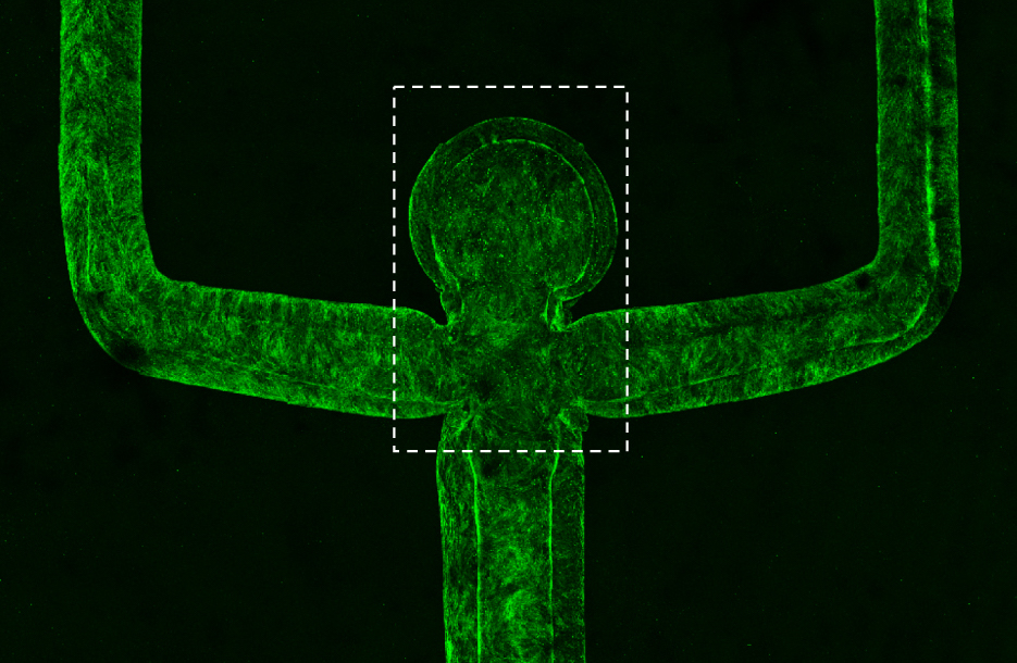
The LLNL-team 3D printed their additive aneurysm’s blood vessels using a sacrificial bio-ink made out of a Poloxamer 407 copolymer and surrounded it with a protein-based hydrogel in the shape of a dome. After printing, the system was cooled to just 4oC, allowing the ink to dissolve, and leaving the team’s vascular structure behind.
“This is an ideal platform, because we can make these flow measurements that would be incredibly difficult to make if you were doing this in an animal.”
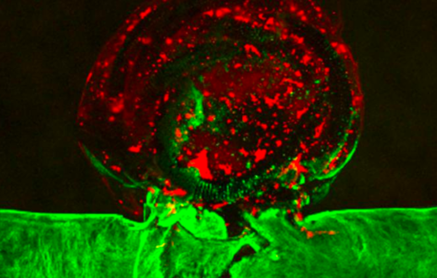
Human brain cells were subsequently introduced to “coat” the model, and cultured for nine days to allow both the vessels and aneurysm to grow. The researchers then deployed two coils using a microcatheter insertion technique, placing the tip of the device at the neck of the aneurysm, and activating it using an electrolytic push wire.
Utilizing a microscopy-based approach, the team were able to observe the procedure in real-time, and noted that the endothelium had began to heal itself shortly after the tests. In future, the researchers believe that their 3D printing approach could be combined with computational modelling, to allow surgeons to perform “test-runs” before performing surgery on a real patient.
“What’s exciting, is that this platform mimics blood vessel compliance and the mechanical stiffness of brain tissue,” concluded Moya. “This makes it ideal to be used as a training platform for surgeons, or as an in vitro testing system for embolization devices.”
Enhancing cerebral understanding via 3D printing
The brain is the most complex organ in the human body, and treating cerebral illnesses remains challenging, but recent bioprinting advances have increasingly provided insights into more effective treatments.
A team of researchers from Tsinghua University have 3D bioprinted brain-like tissue structures that are capable of nurturing neural cells. The cerebral networks were successfully integrated into a lab rat’s brain, and showed great potential as a means of testing drugs.
Similarly, scientists from Oxford University and the Chinese University of Hong Kong, have created a novel bioprinting method, that’s allowed them to better understand how the human brain develops. The team’s new process saw them 3D print human cortical cells into a soft, biocompatible ECM matrigel in both natural and unnatural cell designs.
Elsewhere, researchers at Seoul National University Hospital and the Pohang University of Science and Technology, have bioprinted “glioblastoma-on-a-chip” devices to better understand cancer cells. The team’s cell-based contraptions allowed them to create a predictive model for determining the effectiveness of certain glioblastoma drugs.
The researchers’ findings are detailed in their paper titled “Three-dimensional bioprinting of aneurysm-bearing tissue structure for endovascular deployment of embolization coils.” The study was authored by Lindy K Jang, Javier A Alvarado, Marianna Pepona, Elisa M Wasson, Landon D Nash, Jason M Ortega, Amanda Randles, Duncan J Maitland, Monica L Moya and William F Hynes.
To stay up to date with the latest 3D printing news, don’t forget to subscribe to the 3D Printing Industry newsletter or follow us on Twitter or liking our page on Facebook.
Looking for a new podcast? Be sure to subscribe to the Another Dimension podcast on your chosen podcast player to make sure you never miss an episode.
Are you looking for a job in the additive manufacturing industry? Visit 3D Printing Jobs for a selection of roles in the industry.
Featured image shows the LLNL-led team’s 3D printed aneurysm. Image via LLNL.
