Technology from BIOMODEX, a provider of 3D printed anatomical models, has been used in the simulation of a full cardiovascular procedure to simulate left atrial appendage occlusion (LAAO) – a surgery that aims to prevent blood clot formation in patients suffering from atrial fibrillation.
Physicians at the Montreal Heart Institute in Montreal rehearsed the LAAO procedure for the first time in North America using a bio-realistic haptic simulator from BIOMODEX that contains a 3D printed replica of the patient’s heart.
The successful simulation has validated the company’s technology for its ability to reduce surgical errors and improve the outcomes of procedures.
“Rehearsing a specific case was very helpful – especially one in which we used innovative technologies like the new Abbott Steerable Delivery Sheath for the Amplatzer Amulet occluder, and the transseptal VersaCross Baylis System,” said Dr. Reda Ibrahim, Interventional Cardiologist at the Montreal Heart Institute.
“This realistic simulation allowed us to maximize procedural success and safety.”
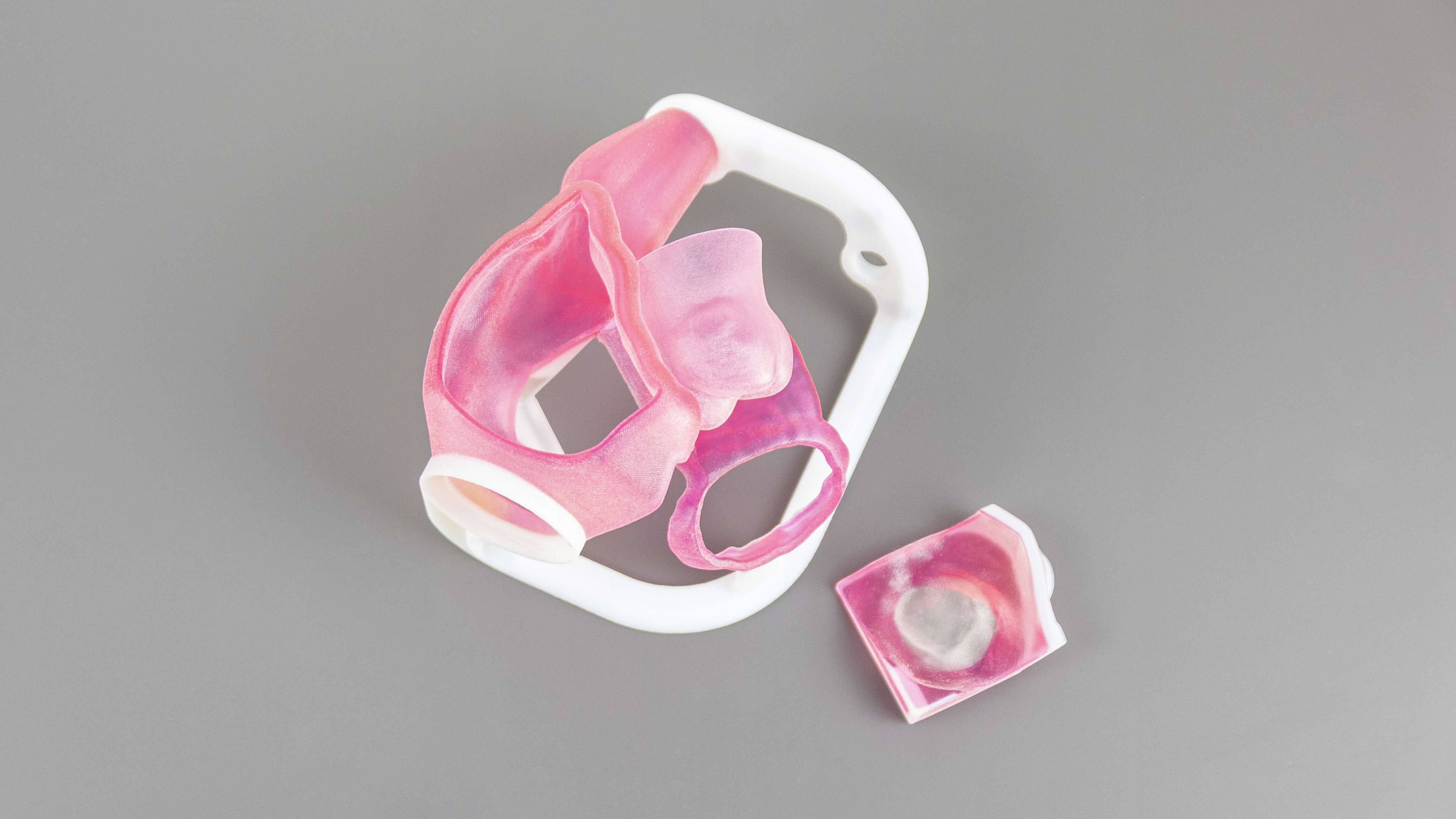
BIOMODEX’s 3D printed heart models
BIOMODEX was founded in 2014 and 3D prints replicas of patient anatomy derived directly from patient-specific medical images such as MRIs and CT scans. The 3D printed models imitate the haptic feedback, texture and feel of real human anatomies, aiding surgeons in validating their medical devices and rehearsing their surgical procedures.
In 2019, the company opened a new US corporate headquarters in Quincy, Massachusetts, containing a 3D printing lab from which the firm could manufacture its anatomical replicas for use in the medical sector. From this, BIOMODEX aimed to distribute the 3D printed anatomical replicas to various physicians, health systems, researchers and medical device makers across North America.
At the start of this year, BIOMODEX launched a new 3D printed training system for the transseptal puncture procedure, which provides a direct route to the left atrium through the intra-atrial septum that separates the left from the right atrium. By emulating the feel, geometry, and haptic feedback of real heart tissue, the model enables cardiologists to practice in a more realistic environment and receive more specialized training.
BIOMODEX’s LAACS technology
Cardiovascular transcatheter therapies have helped to expand the treatment options for patients with structural heart disease. Biomodex has successfully commercialized several products for neurovascular and structural heart applications and is fast expanding its product portfolio.
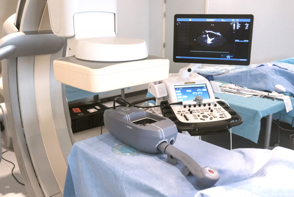
The company’s LAACS technology was used to perform a simulation of an LAAO procedure for the first time using a bio-realistic haptic simulator from BIOMODEX. The breakthrough was achieved by Ibrahim, electrophysiologist Dr. Blandine Mondesert, and cardiac surgeon Dr. Walid Ben Ali, who utilized a 3D printed replica of the patient’s heart. This replica forms part of a novel simulator that provides realistic catheter navigation, haptic feedback, transesophageal echography, and fluoroscopic imaging.
Based on CTA images from the patient, BIOMODEX 3D printed a cardiac anatomy from advanced materials that simulates the biomechanical characteristics and haptic feedback of the heart. The LAACS technology replicates the organ’s blood flow and viscosity, and is also ultrasound compatible. In addition to boosting the confidence of surgeons, the rehearsal is intended to help reduce operating times and decrease the risk of complications during surgery.
“Rehearsing these novel approaches using a patient-specific anatomical model is intended to better prepare physicians and potentially improve patient outcomes,” said Ziad Rouag, President and CEO of BIOMODEX. “It was an honor to support Dr. Ibrahim and his team.”
The rehearsal and live patient procedure were performed and recorded at the Montreal Heart Institute, ahead of Ibrahim and his team presenting their live case at the TVT Structural Heart Summit in Miami, Florida, on July 22nd.
3D printed anatomical models
Producing realistic and accurate representations of the morphology of the human body is vital in the teaching of anatomical knowledge, and as such the 3D printing of such models for educational purposes is continually improving.
In the past year 3D printer OEM Stratasys has provided a software update for its J750 Digital Anatomy 3D printer to better replicate porous bone structures, fibrotic tissue, and ligaments, and medical device manufacturer 3D LifePrints gained ISO certification for the quality assurance procedures behind its 3D printed surgical guides and anatomical models.
Elsewhere, US researchers have evaluated the accuracy of 3D printed medical models produced on a Formlabs Form 3B 3D printer which is used extensively in medical institutions for dental, surgical planning, and educational applications. Most recently, the University of Florence leveraged a 3DUJ-553 3D printer from Mimaki, a manufacturer of wide-format inkjet printers and cutters, to produce 3D printed anatomical models with a previously unattainable degree of color fidelity.
Nominations for the 2021 3D Printing Industry Awards are now open, have your say who is leading the industry now.
Subscribe to the 3D Printing Industry newsletter for the latest news in additive manufacturing. You can also stay connected by following us on Twitter and liking us on Facebook.
Looking for a career in additive manufacturing? Visit 3D Printing Jobs for a selection of roles in the industry.
Subscribe to our YouTube channel for the latest 3D printing video shorts, reviews and webinar replays.
Featured image shows BIOMODEX’s LAACS technology. Photo via BIOMODEX.