The capabilities of a 3DUJ-553 3D printer from Mimaki, a manufacturer of wide-format inkjet printers and cutters, have been leveraged by the University of Florence and Mimaki’s exclusive Italian importer Bompan to produce 3D printed anatomical models with a previously unattainable degree of color fidelity.
During the project, a team of visionary doctors and researchers came together to realize the full color and photorealism capabilities of Mimaki’s 3D printing technology, which they believe could mark a turning point in anatomical models and the teaching of knowledge around the human body.
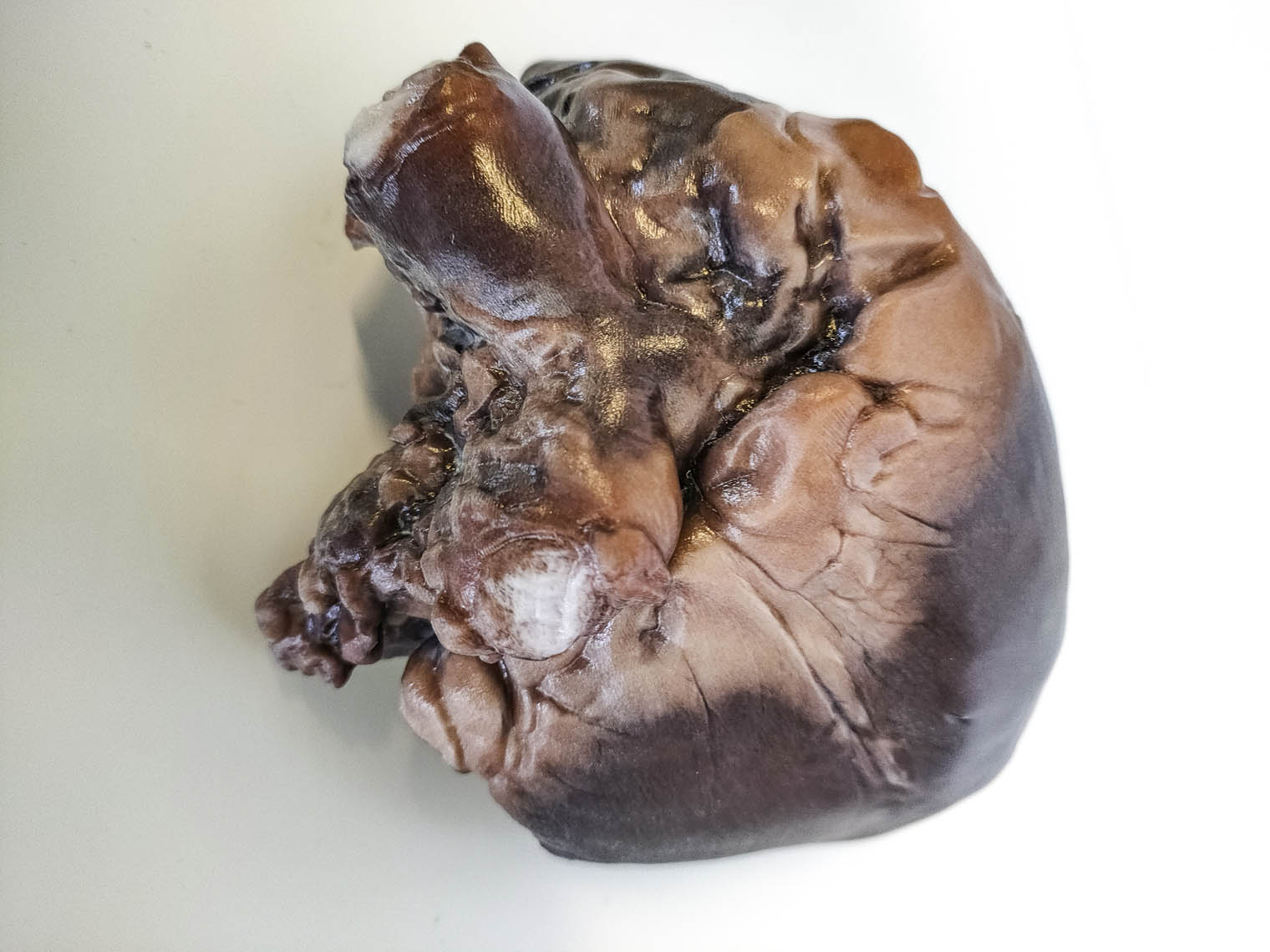
After creating and patenting a unique image implementation and integration algorithm to generate precise graphic reproductions of anatomical systems, the researchers turned to Mimaki’s 3DUJ-553 to print the first model, a heart, with good dimensions, detail definition, and “excellent” color fidelity.
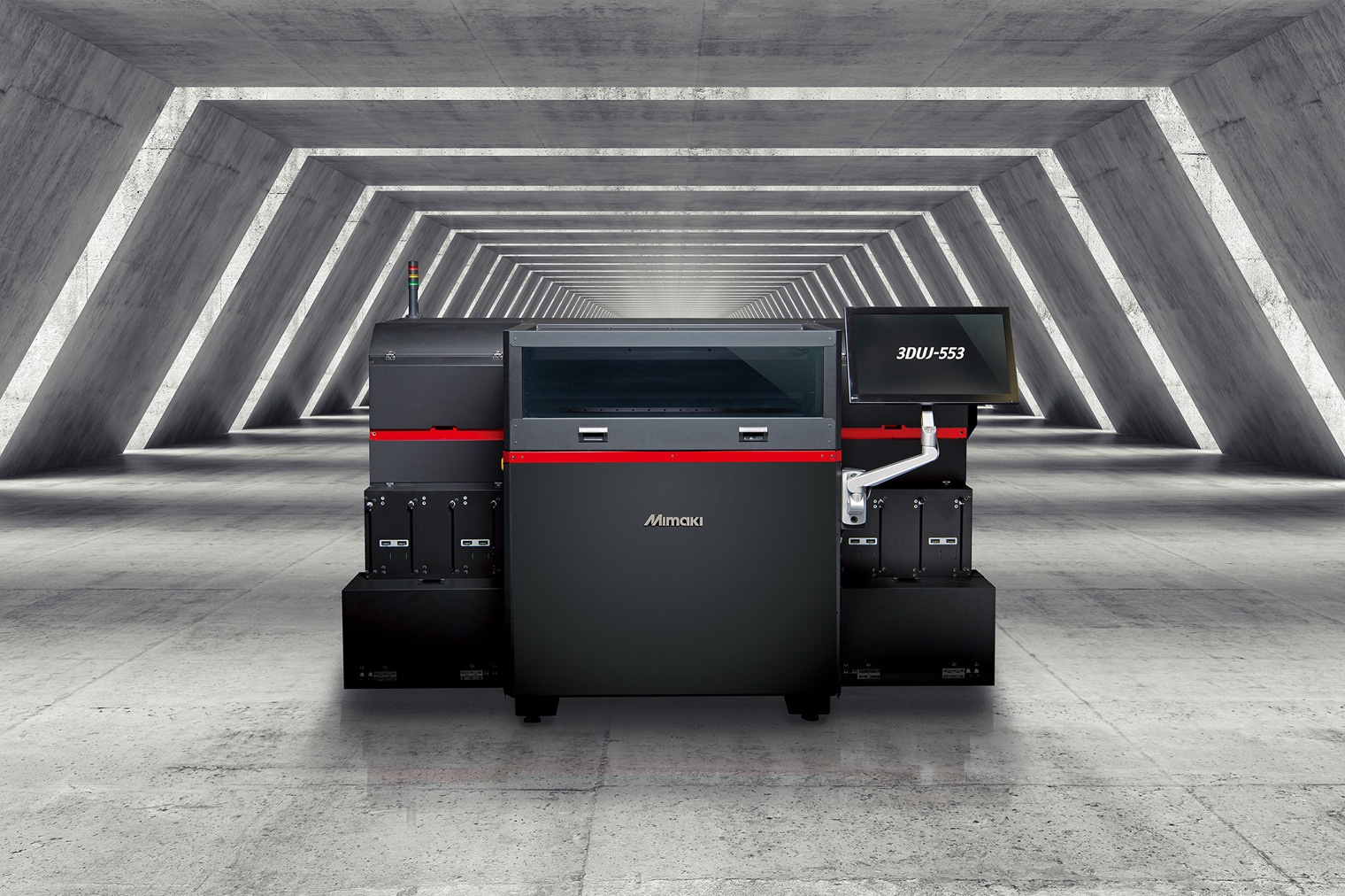
Mimaki’s 3DUJ-553
Mimaki launched its UV LED 3DUJ-553 full color 3D printer in 2017, bringing 10 million possible color combinations to the then mainly monochrome 3D printer market.
The industrial-sized machine is based on the company’s UV inkjet technology which combines a traditional inkjet print head with an economical UV-LED light source. The printer works by jetting successive layers of ink which are instantly cured to build up a full color 3D object.
The 3DUJ-553 has previously been utilized to support the development of engaging exhibits and to produce 3D printed models for public programs at The Smithsonian Exhibit studios in Landover. The machine has also been deployed within the gaming sector to 3D print an intricate model of the winning design of a competition run by Minecraft, the popular sandbox video game created by Swedish developer Mojang.
Since the launch of the 3DUJ-553, Mimaki has identified a need for a smaller entry-level machine on the market at a more affordable price point. The company subsequently launched its full color desktop-sized 3DUJ-2207 3D printer in November last year, which is commercially available for less than €40,000.
The role of 3D printing in anatomical study
For centuries, anatomy has been the basis of medical and scientific knowledge, and remains a vital tool in the training and professional development of doctors around the world. Producing realistic and accurate representations of the morphology of the human body is therefore vital in the teaching of anatomical knowledge, and this is where the benefits of 3D printing technologies can play a key role.
The main method of anatomical investigation is still currently dissection, which allows researchers to gain knowledge and produce an iconographic representation of the structures of the human body. However, due to issues such as the perishable nature of corpses and legal complications, this method has become increasingly less practical.
As such, University of Florence physician and associate professor Ferdinando Paternostro, and doctor Giacomo Gelati, embarked upon finding new ways to fill the existing gap in anatomical investigation.
“During my medical studies, thanks to Ferdinando, who was my professor, I become passionate about anatomy,” said Gelati. “However, I soon became aware of a series of difficulties. Not only was it impossible to take part in anatomical dissection practice sessions, but the iconography used (albeit based on photographs that showed realistic anatomical details and watercolors of great artistic value, despite relying on a schematic representation of anatomical structures) remained flat and static, and therefore not very usable.
“For people who study anatomy or practice medicine, the opportunity to see the real light and colors of an anatomical specimen, and to test its consistency and relationship with the surrounding structures, can really make a difference.”
Leveraging Mimaki’s 3D printing technology
To solve this problem, Gelati created and patented a unique image implementation and integration algorithm that generates a precise graphic reproduction of anatomical systems. Once this had been achieved, the doctors looked to 3D printing to bring their vision to life.
“We had beautiful and effective graphic images available, so we began to consider the opportunity to transform them into 3D, manageable and imperishable objects,” continued Gelati. “We immediately thought about 3D printing, focusing on color fidelity, which is a crucial element for us.”
The researchers turned to Bompan and Mimaki’s 3D printing technology to produce the models, with the technology chosen specifically for its full color capabilities.
“It was a challenge within a challenge,” said Gelati. “Graphic quality and print quality go hand in hand: without a good source file there will never be a good 3D printed object and, conversely, without a good 3D printed, a source file will lose its quality in the printing process. This is why we decided to team up with an excellent 3D color printing company.”
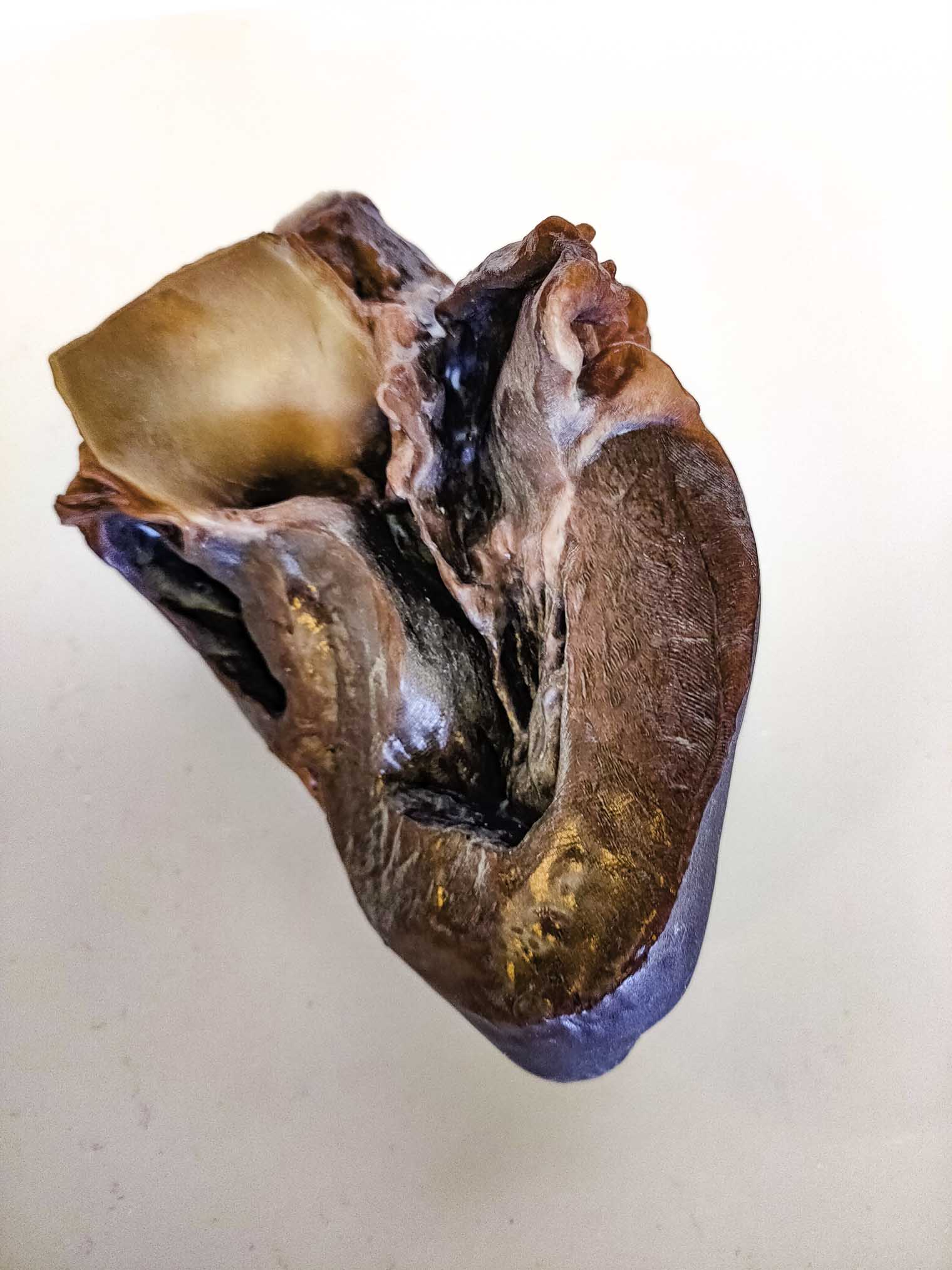
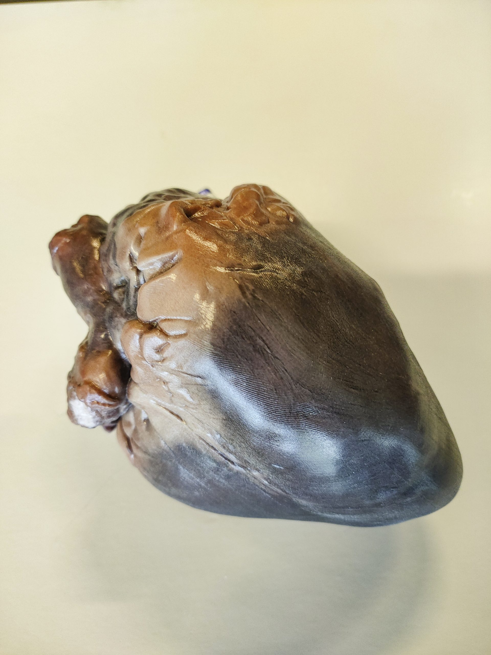
The University of Florence researchers and Bompan worked together to 3D print the first organ model – a heart – using Mimaki’s 3DUJ-553 3D printer. The pilot project proved a success, enabling the creation of a 3D heart model with good dimensions, detail definition, and excellent color fidelity. The ability to reproduce the color quality of the graphic images was the most important factor in the researchers’ choice of technology, according to Paternostro.
“The various anatomical structures we come across when performing surgery or in the dissection room have their own specific color and are surrounded by a topographical context of various different colors,” he explained. “Distinguishing structures in their anatomical topographical context is not particularly easy, and color plays a crucial role.”
As such, the team’s goal is to fully leverage the color quality, replicability, and durability of 3D printed objects within medical practice, to aid the recognition of structures during surgical and anatomical dissections.
“The objects we make by combining our algorithm with Mimaki’s 3D printing technology are chromatically and morphologically realistic, measurable, repeatable,” Paternostro added. “Using this method, we could potentially cross further frontiers. In pathological anatomy, for example, we will be able to create a 3D organ that shows the anomalies caused by a specific disease – thus providing a very useful tool for preparing any surgical intervention and for communicating with the patient.”
Transforming medical-scientific research
Andrea Ferrante, 3D printing specialist, at Bompan believes the project demonstrates the superior qualities of Mimaki’s 3D printing technology and its capability to achieve the color fidelity and ultra-realistic definition of details required for anatomical models, commenting: “We are convinced that this technology will be widely used in a variety of different fields and the opportunities will be boosted by the imminent arrival of the 3DUJ-2207 – a version with a more compact and more accessible design but equipped with the same technology as the 3DUJ-553.”
According to Paternostro, 3D printing generally has the potential to significantly aid in the transformation of teaching, education and medical-scientific research in the future.
“We are still at an early stage, but we are confident that we are on the right track,” he said. “We have the opportunity to replace anatomical models and plastinated anatomical parts – both of great value, but delicate, perishable, and therefore only usable in certain contexts – with 3D printed anatomical pieces, available to universities, research institutes, hospitals and clinics.
“A beautiful anatomical model is functional to learning, communication, medical and surgical practice.”
Subscribe to the 3D Printing Industry newsletter for the latest news in additive manufacturing. You can also stay connected by following us on Twitter and liking us on Facebook.
Looking for a career in additive manufacturing? Visit 3D Printing Jobs for a selection of roles in the industry.
Subscribe to our YouTube channel for the latest 3D printing video shorts, reviews and webinar replays.
Featured image shows the University of Florence and Bompan worked together to 3D print a heart using the Mimaki 3D UV LED 3DUJ-553 printer. Photo via Mimaki.