
Researchers at IFW Dresden and Chemnitz University of Technology have 3D printed a microtube that can be used for guiding sperm to treat cancer.
Naturally adapted for survival in the female reproductive system, the drug-loaded sperm cells provide an intuitive treatment for gynecologic cancers that, in the U.S. alone, affect more than 100,000 women each year.
The ideal partner for controlled drug delivery
In addition to a natural ability to last inside a uterus, sperm exhibit a number of other qualities favourable for controlled drug delivery. Self-propulsion, and an ability to pierce a cell membrane, can be utilized to deliver drugs to the very center of cancer cell where it is most effective. Additionally, with a “paddle-like” shape, the cells have a high drug loading capacity.
3D printed smart devices for sperm
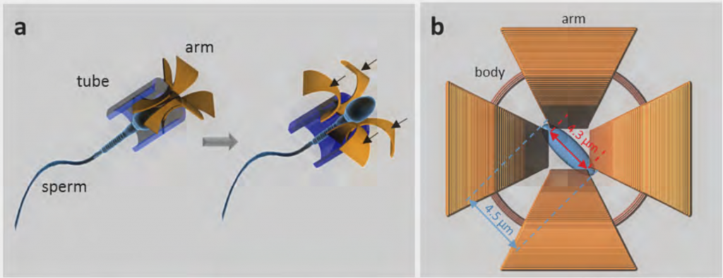
Making a smart device out of sperm cells is done by 3D printing on a nanoscopic scale (3D nanolithography). To enable control and maximize the natural properties of sperm cells, researchers created a minute device capable of releasing the sperm upon contact with another substantial structure.
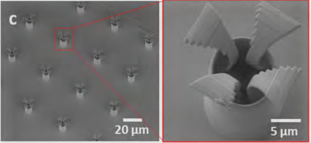
The device takes the form of a “tetrapod”, with four arms that bend on contact with another cell wall, allowing a sperm to swim freely out of the casing and into a cancerous cell.
Before introduction to the tetrapod, the sperm is soaked with a form of Doxorubicin medication, specifically developed for the treatment of gynecological cancers. The tetrapods were made from light-reactive IP-Dip material from Nanoscribe GmbH, and then coated in iron to make them magnetic.
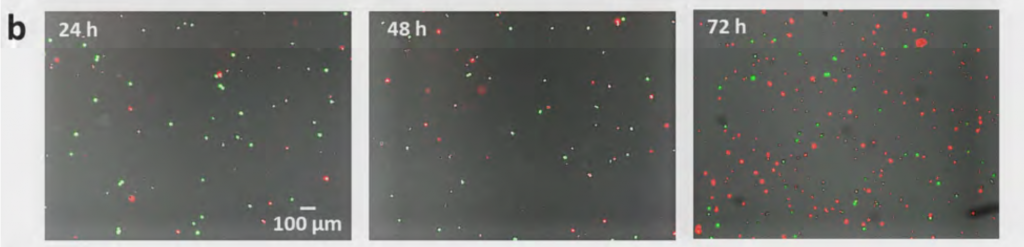
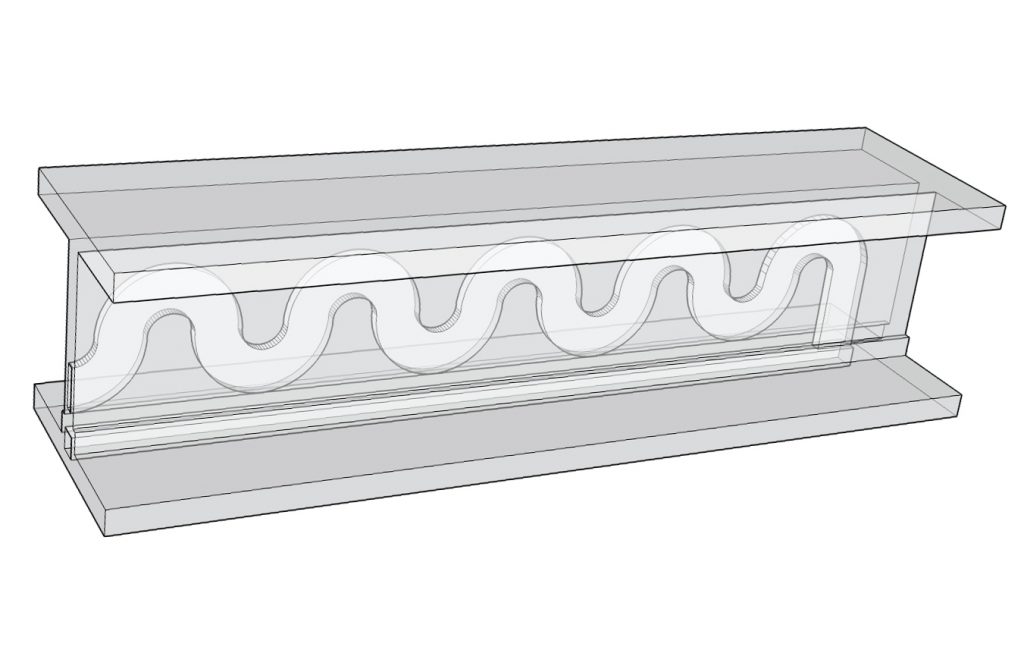
The tetrapod sperm, along with immortal HeLa cervical cancer cells, were then introduced to a microfluidic device to study the effects of contact between the cells. Results showed “more than 2/3 (15 out of 22) of the coupled motors were shown to successfully release sperm cells.”
Conclusions and further research
Though there are still come challenges to overcome before these methods can be tested inside a living organism, conclusions state that “sperm-hybrid systems may be envisioned to be applied in in situ cancer diagnosis and treatment in the near future.”
As chemotherapy drugs cause damage to living cells, and are used to treat such a volatile and still incomprehensible condition, the need for making controlled drug delivery devices is especially pertinent for cancer treatments. Other research looking to create such devices include a watch-inspired mechanism from Columbia University, and the University of Illinois Urbana-Champaign’s 3D printed “bio-bots”.
Microfluidic devices are also key in such research. These devices provide a small-scale test environment with channels that more accurately represent vessel structure inside the body. With the help of 3D printing, the Lewis Lab at Harvard University were able to replicate a kidney vessel structure on a microfluidic chip, and there are many projects looking to encourage the adoption of microfludic study in labs.
Sperm-hybrid micromotor for drug delivery in the female reproductive tract is published open access via Cornell University Library. It was co-authored by Haifeng Xu, Mariana Medina Sanchez, Veronika Magdanz, Lukas Schwarz, Franziska Hebenstreit and Oliver G. Schmidt.
For more of the latest 3D printing medical research sign up to the 3D Printing Industry newsletter and follow our active social media channels.
Don’t forget to vote now in the first annual 3D Printing Industry Awards.
Featured image shows sperm under a microscope. Photo by Iqbal Osman on Flickr
