Since the days of “be seen and not heard” in education, teachers have learned that learning is an interactive, dialectic process. Students all have a variety of methods by which they learn; seeing a lesson on a chalkboard may not be enough for kinetic learners. And, when it comes to STEM education, there’s something about being able to physically grasp a scientific or mathematical model that makes a concept that much easier to understand. 3D printing has pushed the capability of kinetic education, allowing for the construction of elaborate models and, in the field of medicine, 3D printed models could mean saving lives.
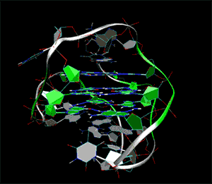
At the University of Alabama’s 3D Printing Studio, the school’s science and engineering librarian, Dr. Vincent F. Scalfani, worked with researchers at the School of Pharmacy at University College in London, Drs. Stephan A. Ohnmacht and Stephen Neidle, to transform lab X-rays into digital 3D models. By converting X-ray crystallography information of the cancer-inducing G-quadruplex molecule in addition to the drug targeting it into 3D digital models, the team hopes to 3D print representations of the molecules for research and education purposes.
Already, the 3D printed G-quadruplex DNA is helping researchers and students increase their understanding of the cancer and how to treat it. In fact, it’s in the process of being used in preclinical trials for studying pancreatic cancer. Dr. Scalfani explains, “Preparing the G-qaudruplex DNA sequence for 3D printing was a challenge and certainly pushed the limits of what we thought was possible in the UA 3D Lab. The structure is extremely intricate, containing multiple areas of stacked functional groups (the quadruplex) that are all surrounded by a common outer loop (the DNA backbone).The 3D printed G-quadruplex is stunning; you can see all of the symmetry, facets and angles within the molecule.”
Drs. Ohnmacht and Neidle point out how the handheld model immensely improves upon former 2D images, with Ohnmacht saying, “Having a live model is invaluable; visualizing distances of bonds, electrostatic interactions and angles is easy and allows for further optimization of these anti-cancer molecules. The printed 3D model is actually a real molecular structure that has been designed, synthesized and then crystallized in the London labs. G- quadruplex DNA is unusual as it is four-stranded, not two stranded like ‘normal’ double helical DNA we know.”
This work, I think, can only be complemented with ongoing research into virtual reality. Though an immersive virtual, holodeck-type experience may give doctors the ability to zoom in and explore 3D models, a 3D printed tactile aid can only improve one’s understanding of such a model. After all, we evolved from apes, so there’s still a lot of learning left in our hands and feet.
Source: Phys.org