Hearing aids are one of the earliest examples of the added value 3D printing can bring to a commercial market. Coupled with 3D scanning technology, additive manufacturing has made it possible for companies like Sonova to improve the quality of existing products through mass customization.
Recently presenting at the Radiological Society of North America (RSNA), Dr. Jeffrey D. Hirsch and colleagues from the University of Maryland School of Medicine (UMSOM) have demonstrated the next step in custom 3D printed aural therapy.
The risk of aural implants
Hirsch et al.’s research at UMSOM concerns radiology and the ossicles of the middle ear. Composed of three small bones, commonly known as the hammer, anvil, and stirrup, the ossicles create hearing loss when damaged through trauma or infection.
In these cases, hearing can be restored using prosthetic struts that restore bones to their correct places. However, these procedures all-too-often result in failure.
Dr. Hirsch explains, “The ossicles are very small structures, and one reason the surgery has a high failure rate is thought to be due to incorrect sizing of the prostheses,”
“If you could custom-design a prosthesis with a more exact fit, then the procedure should have a higher rate of success.”
A snap fit
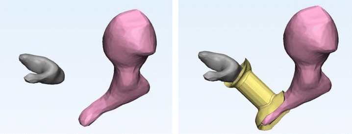
Using cadaver CT scan data, Hirsch et al. have developed a range of customizable prosthetic implants for the middle ear. Each implant intuitively fits with the bone it is meant for, proved in a “blind test” by aural surgeons.
As detailed in the accompanying study, “Four surgeons […] performed insertion of each prosthesis into each middle ear, blinded to the bone from and for which each was designed.”
“The surgeons were asked to match each prosthesis to its correct parent bone.”
Remarkably, all of the surgeons in the experiment matched the prosthesis to the right ossicular bone.
“This study highlights the core strength of 3D printing — the ability to very accurately reproduce anatomic relationships in space to a sub-millimeter level,” said Dr. Hirsch “With these models, it’s almost a snap fit.”
Further reading
3D printed implants are commonly used in operations on the spine, skull and in dentistry. The next step for Hirsch at al. is to make implants for the ossicles using biocompatible matierals, and investigate the benefits of porosity for regrowth under the skin.
Surgical reconstruction of the ossicular chain with custom 3D printed ossicular prosthesis was co-authored by Jeffrey D. Hirsch, Richard L. Vincent and David J. Eisenman. It is published online, open access, in the journal 3D Printing in Medicine.
Stay up to date with the latest 3D printing research – sign up to the 3D Printing Industry newsletter, follow us on Twitter, and like us on Facebook.
Get involved with 3D printing events near you here.
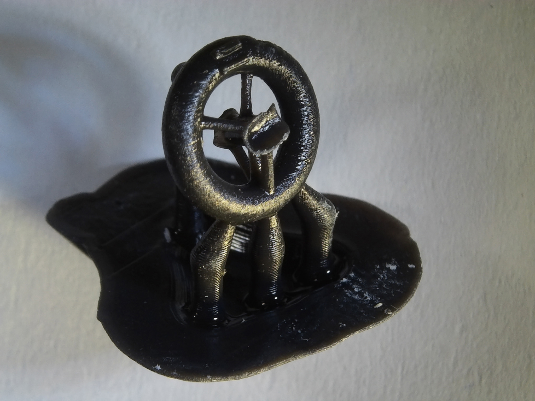
Featured image shows 3D printed bone implants from the University of Maryland School of Medicine with penny for scale. Photo via RSNA
