3D printing may hold the key to beating the primary cause of death in the U.S. – heart disease. With almost a quarter of deaths in North America attributable to heart disease, this new research from the University of Minnesota has far reaching implications.
Brenda Ogle, an associate professor of biomedical engineering at the University of Minnesota, described the work a, “significant step forward.” The professor added,
We feel that we could scale this up to repair hearts of larger animals and possibly even humans within the next several years.
As we’ve previously reported 3D printing is finding increasing application for the treatment of cardiac patients.
A digital model of proteins
Starting with a digital model of the proteins that compose heart tissue, 3D printing was used to develop the digital heart patch.
The team of researchers used, “multiphoton-excited 3D printing to generate a native-like extracellular matrix scaffold with submicron resolution.”
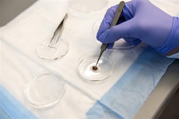
The scaffold was then seeded with cardiomyocytes, smooth muscle cells, and endothelial cells, to “generate a human-induced pluripotent stem cell–derived cardiac muscle patch (hCMP).”
A study of the 3D bioprinted cell patch using mice found a, “significant increase in functional capacity” of their hearts after an induced heart attack. In the month following the heart attack the bioprinted heart patch was absorbed by the animal.
3D Printing and photochemistry
Published in Circulation Research the paper describes how the, “principles of 3D printing and photochemistry can be combined to generate an ECM scaffold with unprecedented resolution from a template based on the architecture of native myocardial ECM.”
Professor Ogle said, “Given the complexity of the heart, we were encouraged to see that the cells had aligned in the scaffold and showed a continuous wave of electrical signal that moved across the patch.”
The researchers say the use of 3D printing permitted generation of the scaffolds with “exceptional resolution.” In concluding the research the scientists say that such hCMP’s may, “significantly improve recovery from ischemic myocardial injury.”
Repairing larger hearts with 3D printing
The next stage of the research will, “scale this up to repair hearts of larger animals and possibly even humans within the next several years,” said Ogle. This phase of the research will create patches that can be used on pig hearts, before moving to human trials.
Ling Gao, Molly E. Kupfer, Jangwook P. Jung, Libang Yang, Patrick Zhang, Yong Da Sie, Quyen Tran, Visar Ajeti, Brian T. Freeman, Vladimir G. Fast, Paul J. Campagnola, Brenda M. Ogle and Jianyi Zhang worked on the research, which is published in full here.
For all the latest 3D printing news, subscribe to our free newsletter here and follow our active social media accounts.
If you think this research should receive a 3D Printing Industry Award, then vote now in the 1st Annual 3D Printing Industry Awards.
