Researchers at the Massachusetts Institute of Technology (MIT) have developed a method for 3D printing a flexible and soft replica of a patient’s heart.
The team can then direct the activity of the replica to imitate the patient’s blood-pumping capabilities. With this custom robotic heart, the team hopes to assist doctors in tailoring treatments to patients’ heart-related forms and functions. The soft robotic models are patient-specific and may aid clinicians in choosing the ideal implant for a particular patient.
“All hearts are different. There are massive variations, especially when patients are sick. The advantage of our system is that we can recreate not just the form of a patient’s heart, but also its function in both physiology and disease,” said Luca Rosalia, a student in the MIT-Harvard Program in Health Sciences and Technology.
Mechanical Engineering Professor Ellen Roche said, “Being able to match the patient’s flows and pressures was very encouraging. We’re not only printing the heart’s anatomy but also replicating its mechanics and physiology. That’s the part that we get excited about.”
What is the procedure for this innovative approach?
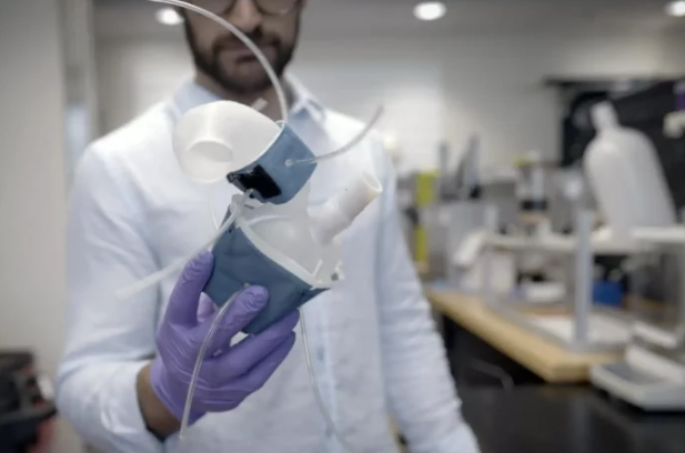
The method entails transforming medical images of a patient’s heart into a 3D computerized model, which the team then 3D printed with polymer-based ink. This produces a soft, flexible shell that is precisely the structure of the patient’s own heart. This method additionally has the potential to print a patient’s aorta, which is the main artery that transports blood from the heart to the remainder of the human body.
The team created sleeves similar to blood pressure cuffs that encase around a 3D printed heart and aorta to imitate the heart’s pumping action. Researchers can further tune the outflowing air to rhythmically pump up the sleeve’s bubbles and contract the heart to imitate its pumping activity when the sleeve is bonded to a pneumatic system.
The researchers can also compress the vessel by inflating an independent sleeve surrounding a 3D printed aorta. The team claims that this narrowing can be tuned to imitate aortic stenosis, an ailment in which the aortic valve narrows, requiring the heart to exert more effort to force the blood to circulate through the body.
Aortic stenosis is generally treated by surgically inserting a synthetic valve intended to enlarge the natural valve of the aorta. According to the researchers, in the future, doctors could employ the process to 3D print a patient’s heart and aorta, then implant a multitude of valves into the printed model to determine which design offers the ideal function and is fit for the patient.
In their study, the researchers utilized medical scans from 15 patients with aortic stenosis. Each patient’s images were converted into a 3D computer model of the patient’s left ventricle (the primary pumping heart chamber) and aorta. The model was then loaded into a 3D printer, which produced a soft, anatomically correct shell that resembled the ventricle and vessel.
“There is a lot of interest in the medical field in using 3D printing technology to accurately recreate patient anatomy for use in preprocedural planning and training,” said Wang, a vascular surgery resident at Beth Israel Deaconess Medical Center in Boston.
The potential of 3D printing organs
While MIT’s replica heart is not intended to be implanted into a patient, other 3D printing firms are actively exploring such applications – commonly referred to as regenerative medicine.
In the next five years, 3D Systems and United Therapeutics hope to begin human clinical trials for its personalized 3D printed lung scaffolds, possibly bringing the objective of fully transplantable 3D printed organs one step closer. United Therapeutics, a biotechnology company, demonstrated what it calls the “world’s most complex 3D printed object” in collaboration with 3D printer manufacturer 3D Systems at the LIFE ITSELF conference in San Diego. The project has so far created a 3D printed human lung scaffold that can demonstrate gas exchange in animal models and has further intended to cellularize the scaffold with a patient’s own stem cells to generate palatable, transplantable human lungs.
3D Systems says the 3D printed lung scaffold design contains 44 trillion voxels that lay 4,000 km of pulmonary capillaries and 200 million alveoli. For several years, both companies have collaborated on advancing their Print to Perfusion process, which is intended to enable the 3D printing of high-resolution scaffolds that can be perfused with living cells to generate tissues.
“It was exciting to show the public our 3D printed human lung scaffold, but we’re thrilled to share that our 3D printed lung scaffolds are now demonstrating gas exchange in animal models,” said Dr. Martine Rothblatt, United Therapeutics’ Chairperson and CEO. “We are regularly printing lung scaffolds as accurately as driving across the United States and not deviating from a course by more than the width of a human hair.”
Elsewhere, a Utrecht University research team successfully created working livers employing an ultrafast volumetric 3D bioprinting method. The volumetric bioprinting method used visible light tomography to successfully 3D print miniature stem cell components by creating “transparent” cells which implied they preserved their resolution and capacity to carry out biological processes. The liver units, printed in less than 20 seconds, could imitate key toxin elimination processes that natural livers conduct in our bodies, potentially opening up new avenues for regenerative medicine and personalized drug testing.
Additionally, Matricelf, an Israeli regenerative medicine firm, signed an exclusive global licensing agreement for a patent-pending 3D bioprinting technology with Ramot, a Tel Aviv University (TAU) technology transfer firm. The technique, as per Globes, utilizes stem cells and extracellular matrix (ECM) from a patient to bioprint implants for the regrowth of injured organs and tissues in the body. The method was in the patent process for approval in the United States and Europe in 2022.
What does the future of 3D printing for the next ten years hold?
What engineering challenges will need to be tackled in the additive manufacturing sector in the coming decade?
To stay up to date with the latest 3D printing news, don’t forget to subscribe to the 3D Printing Industry newsletter or follow us on Twitter, or like our page on Facebook.
While you’re here, why not subscribe to our Youtube channel? Featuring discussion, debriefs, video shorts, and webinar replays.
Are you looking for a job in the additive manufacturing industry? Visit 3D Printing Jobs for a selection of roles in the industry.
Featured image shows a 3D printed heart replica. Image via MIT.