
Research headed by bioengineers from Rice University and the University of Washington (UW) has advanced the possibility of bioprinting human organs from living cells and hydrogels.
By adding common food dyes, the hydrogels used in the experiment were made photocurable for DLP printing and suitable for the intended application. Food colors, based on natural products such as turmeric powder and blueberries were identified by the researchers as “nontoxic light blockers”, compared to Sudan I, an organic compound, which is unsuitable for bioapplications.
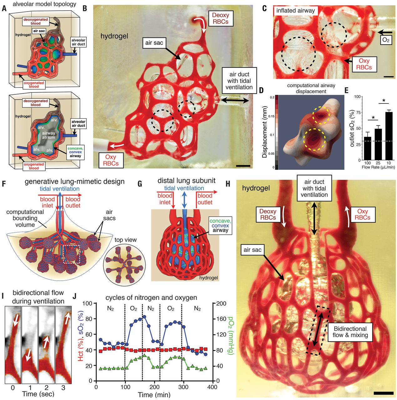
Using non-toxic hydrogels and DLP printing the scientists succeeded in producing a complex network of vessels which are physically and chemically intertwined.
In addition to this, the possibility of transplanting the structure into mice with liver injury was also evaluated. The next step in the research is to make these tissues scalable so they could be transplanted into human bodies.
Mimicking the human lung
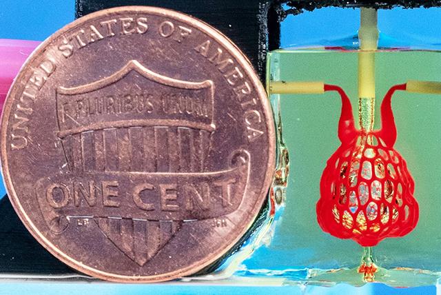
Using living cells, hydrogels, and a DLP printer, an air sack surrounded by multivascular tubes was bioprinted. The organ mimics the behavior of a human lung.
Jordan Miller, assistant professor of bioengineering at Rice’s Brown School of Engineering and one of the leading the researcher, explained, “our organs actually contain independent vascular networks — like the airways and blood vessels of the lung or the bile ducts and blood vessels in the liver. These interpenetrating networks are physically and biochemically entangled, and the architecture itself is intimately related to tissue function.”
“Ours is the first bioprinting technology that addresses the challenge of multivascularization in a direct and comprehensive way.”
Organ replacement
According to the UK’s NHS, there are currently more than 6,000 people waiting for organ transplantation, whereas in the U.S, the figure is nearly 114,000.
One of the primary risks with organ transplantation is that the immune system of the host may reject the transplanted organ, thereby risking the life of the patient.
With the help of bioprinting, organs can be created using the patient’s own cells. This could potentially minimize the risk of rejection of the transplant.
However, for several decades, a major hurdle to progress of bioprinting organs has been the difficulty of creating complex vascular networks inside soft materials in which cells are able to thrive. As Miller explained, “One of the biggest road blocks to generating functional tissue replacements has been our inability to print the complex vasculature that can supply nutrients to densely populated tissues.”
Bioprinting human organs
In the latest research, bioengineers succeeded in creating an air sac surrounded by a complex tubular structure that mimics blood vessels. The pumping of the air sac facilitates the mixing of blood in the surrounding vessels.
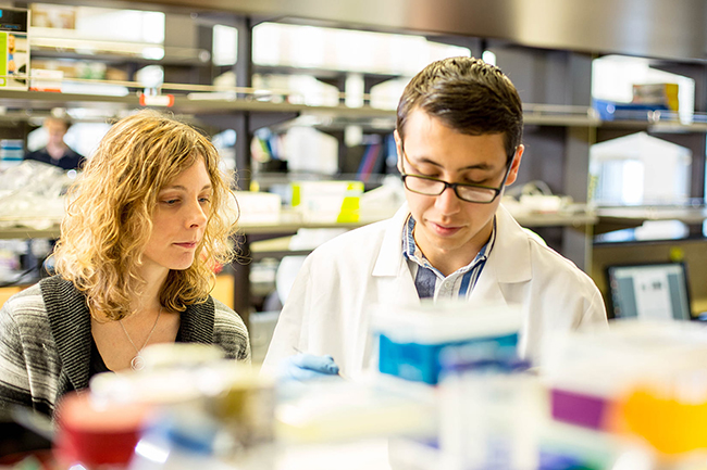
Kelly Steven, a UW assistant professor of bioengineering and research leader said, “With this work we can now better ask, ‘If we can print tissues that look and now even breathe more like the healthy tissues in our bodies, will they also then functionally behave more like those tissues?’ This is an important question, because how well a bioprinted tissue functions will affect how successful it will be as a therapy.”
“The liver is especially interesting because it performs a mind-boggling 500 functions, likely second only to the brain […] The liver’s complexity means there is currently no machine or therapy that can replace all its functions when it fails. Bioprinted human organs might someday supply that therapy.”
A lot of the hardware for research was assisted by open-source 3D printing technology, in particular, the RepRap project, UltiMachine, and Prusa. For the purpose of the study, the research team created an open-source DLP bioprinter called “stereolithography apparatus for tissue engineering,” or SLATE.
Furthermore, Miller and Bagrat Grigoryan, a bioengineering postgraduate from Rice had the idea of adding food dyes to the material which could make the hydrogel absorb blue light and make it photocurable.
The authors of the research state, “We identified candidate photoabsorbers among food additives whose absorbance spectra encompass visible light wavelengths that can be used for biocompatible photopolymerization.”
The design of the air sac was made in collaboration with Nervous System, a Massachusetts-based design studio.
Bioprinting and regenerative medicine
In recent years 3D printing has shed new light on regenerative medicine. Some of the applications of bioprinting reported recently include treatment of scarred lesions and use of stem cells and bioprinting to create tendons and ligaments.
In the coming years, it is expected that bioprinting technology will develop more rapidly, as Miller added, “We envision bioprinting becoming a major component of medicine within the next two decades.”
The research team has made the hydrogel design file publicly available. Miller said, “Making the hydrogel design files available will allow others to explore our efforts here, even if they utilize some future 3D printing technology that doesn’t exist today,”
“We are only at the beginning of our exploration of the architectures found in the human body […] We still have so much more to learn.”
The paper concludes, “We have identified readily available food dyes that can serve as potent photoabsorbers for biocompatible and cytocompatible production of hydrogels containing functional vascular topologies for studies of fluid mixers, valves, intervascular transport, nutrient delivery, and host engraftment.”
“With our stereolithographic process, there is potential for simultaneous and orthogonal control over tissue architecture and biomaterials for the design of regenerative tissues.”
The research discussed in this article is published as Multivascular networks and functional intravascular topologies within biocompatible hydrogels in the journal Science. It was jointly authored by Bagrat Grigoryan, Samantha J. Paulsen, Daniel C. Corbett, Daniel W. Sazer, Chelsea L. Fortin, Alexander J. Zaita, Paul T. Greenfield, Nicholas J. Calafat, John P. Gounley, Anderson H. Ta, Fredrik Johansson, Amanda Randles, Jessica E. Rosenkrantz, Jesse D. Louis-Rosenberg, Peter A. Galie, Kelly R. Stevens, and Jordan S. Miller.
2019 3D Printing Awards are almost here. Vote now in the category of 3D printing for a better world.
To learn more about bioprinting and regenerative medicine subscribe to our 3D printing newsletter. You can also get live updates by following us on Twitter and Facebook.
For jobs in the manufacturing industry, visit our 3D Printing Jobs page.
Featured image shows the bioprinted lung-like air sac. Image via the University of Washington.