With the advantage of high resolution providing facial designs that can be created based on the FACS (facial active coding system), 3D photography scanning and printing of the subject will create images with connected feature grids. This allows the angles of the craniomaxillofacial surface to be observed for specific unique aspects of physical characteristics, for underlying anatomical bone structures based on eye to midline features.
The nasal process provides the centerpiece of anatomical facial mapping and organization which affects how the individual is viewed by the world around them. These connected grids with the aid of imaging allow for facial feature approximation for important craniofacial-facial planning creating vital structures that will be 3 D printed according to accepted innovation in FACS (Facial active coding system) design. Medical grade silicone soft tissue prosthetics with colour are being created out of the UK with the Picsisma printer by Fripp Design as far back as 2013.
Imaging will display facial mapping and preoperative planning for future surgical repair through comparison of previous features and desired outcome reflected in design. With the tissue selection, it will be possible to apply coverage the foundation structure at several different points in order to create the desired look, providing a foundation for nasal process approximation. From the Rhinoplasty gliding imprint, there will be several places or points of insertion (not only for tendons of the face for the zygomaticus major and minor) but allow for characteristics vital to the facial features and function. The 3D print Rhinoplasty system would allow for openings for the passages for contouring across the branches of the facial nerve.
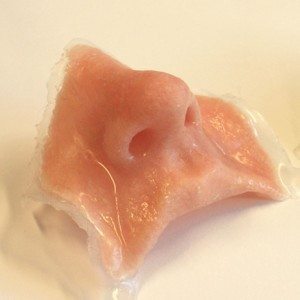
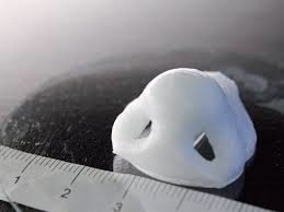
These implants would be made of an FDA approved material consisting of cartilage material to allow for the system to be incorporated into the body’s natural framework.
This new bio-printing technique is being performed with an ETA of 16 minutes by ETH out of Zurich Switzerland with the advantage of autologous cells. The application will apply to both aesthetic and hospital settings involving neck dissection or patients that have complicated work done. The passage of muscle beneath the skin surface that may require rebuilding would involve an initial scan that could create a part to properly fit into place to match the initial impression of the bone structure to overlap any gaps in the connected anatomy.
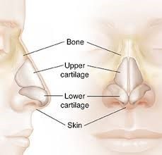
The skin muscle resurfacing allows for the adjustments and fittings to the foundation contour. Several different designs can be created based on the FACS (facial active coding system). 3 D photography of the subject will create images with computer aid design 3 Dimensional software connected grids, allowing the angles of the craniomaxillo-facial surface to be observed for specific unique features and physical characteristics for underlying anatomical bone structures based on eye to midline features in addition to nasal process line.
These connected grids allow for facial feature approximation and craniofacial-facial planning creating a triangular structures of the nasal orbital ethmoid complex that will be important based on accepted FACS (Facial active coding system) to be redesigned. 3 D photography will display features of emphasis for facial mapping and preoperative planning for future surgical repair.
Facial detection will include the discussed prominent and established features which include space between the eyes or central distance including the iris, the depth of the nasal passage, the nasal bridge and protuberance. With the overlapping of demarcating lines, it will be possible to stabilize the foundational structure at several different points and angles in order to create the desired look providing a foundation. Facial Prototyping, which is quickly becoming the standard, has been performed with the 3D systems model 660 PRO by Dr. Avsar with great results out of NYC. These Rhinoplasty implant by Dr. Pablo Prichard out of Phoenix would be made of an FDA approved material to allow for the system to be incorporated into the body’s natural framework.
When evaluating a look at innovations discussed, facial impression with biometric design could be valuable to create the muscles of the orbital wall or even create an individually fitted implant. Optimized imaging equipment will successfully maintain a 3 D visual spatial environment to effectively create a network of design throughout pre-operative, intra-operative, that can be duplicated post-operatively is pivotal objective for the project. Additional outcomes include advantageous visualization and comparison of pre-operative and post-operative key surgical landmarks through datasets/ templates for comprehensive form that define successful design and in correcting specific feature types targeted for enhancement.
Adjustable placement surrounding the foundation of the area of resurfacing that would be replaced with proper alignment surrounding the implant allowing for pressure to be placed along the overlapping belts Once this system has been applied in several settings, then a thoughtful 3 D Computer Aided Design can be created based off successful outcomes.