The use of 3D printed models to help doctors and even medical students prepare for surgeries has been a strong driver of 3D printing adoption; now that 3D printing is becoming more accessible worldwide, it is enabling more surgeons to perform operations that used to be too complex and expensive, especially when delicate areas, such as the brain and the spinal column, are involved.
One recent example comes from India, where local 3D printer manufacturer Divide by Zero used its newest Accucraft i 250+ FDM 3D printer to help JIPMER (Jawaharlaal Institute of Medical Research in Visakhapatnam, India) to plan a surgery on a 4 year old girl whose skull greatly exceeds the average size for her age.
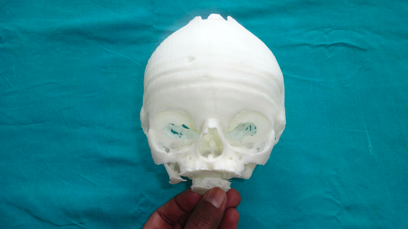
The doctors first captured a CT scan of the girl’s skull, sending the data to Divide by Zero to be converted for 3D printing. The 3D model was then 3D printed with the Accucraft’s 80 micron maximum resolution. The resulting 3D printed model was then used to plan the craniosynostosis for her fronto-orbital advancement. Performing a mock surgery on the 3D printed skull, the surgeons were able to gain awareness of the intraoperative difficulties that could occur and better prepare to deal with them.
Plastic surgeons, and surgeons in general, used to prepare for operations with 2D photographs. The lower costs and wider accessibility of 3D printing technologies have changed everything, making it affordable for many centers, at least for complicated cases. Multicolor and multi material 3D printing capabilities can help even more in complex surgeries, such as the one recently performed at the Centro Médico, Campinas, in São Paulo, Brazil.
Jorge Vicente Lopes da Silva, chief of the Tridimensional Technologies Division of the Center for Information Technology Renato Archer (CTI), has been aiding doctors and surgeons in the creation of 3D models for years. The complex tumor asportation procedure performed at Centro Medico Campinas is one of Silva’s most significant achievements.
The tumor was located in close proximity to their 12 year old patient’s spinal column, close to vital arteries and kidneys, and was considered inoperable; however, Dr. Jose Carlos Barbi Gonçalves was able to complement virtual models with Silva’s 3D printed model to carefully plan the surgery.
The tumor was perfectly removed, along with five of the patient’s vertebrae, which, as we recently had the chance to see, could soon be replaced by perfect 3D printed vertebral implants.