
Researchers from China’s Sichuan University, Belgium’s Ghent University and The University of California San Diego, have developed a method of 3D printing a human ear inside of the body.
The research team’s newly developed Digital Light Processing (DLP)-based technique uses a near-infrared light beam to enable the non-invasive in situ 3D bioprinting of a human ear. This new method could allow doctors to repair human ears that have been damaged by multiple sporting injuries or accidents, and open a new avenue of 3D printing research in non-invasive medicine.
Additive manufacturing tissues using bioprinting
Bioprinting has been utilized to create personalized structures for a range of medical applications in recent years, especially in regenerative medicines. Using inkjet, extrusion, laser direct writing and light-assisted 3D printing techniques, scientists have been able to fabricate living organs and tissues. Many of these in vivo applications require invasive surgical implementation, or in situ 3D printing at the exposed trauma, both of which require exposure of the application site. Moreover, with internal injuries under the skin, surgery which uncovers trauma could damage the surrounding tissues, causing a secondary injury.
The researchers developed an alternative DLP 3D printing process, which enabled them to noni-nvasively fabricate tissue-covered bioinks into customized products, including living tissue constructs in situ. Previous approaches have used the DLP method for multiple-tissue reconstruction or repair, including spinal cord, peripheral nerve and blood vessel injuries. Conventionally, ultraviolet (UV) or blue light is utilized to assist bioprinting via photopolymerization, but these are difficult to use as a tool for non-invasive manufacturing, because of their poor tissue-penetration capacity.
Due to its deep tissue penetration capabilities, the researchers developed a Near-Infrared (NIR) light-based method, instead of using a UV or blue light-based approach. Traditionally used for controlled-drug release in patients, the precise control of the NIR-induced efficient photopolymerization enables the non-invasive fabrication of the tissue-covered bioink into structured products. On the basis of the design of a Digital NIR Photopolymerization (DNP) process, the research team created a non-invasive in vivo 3D bioprinting system.
Using 3D bioprinting to produce a human ear
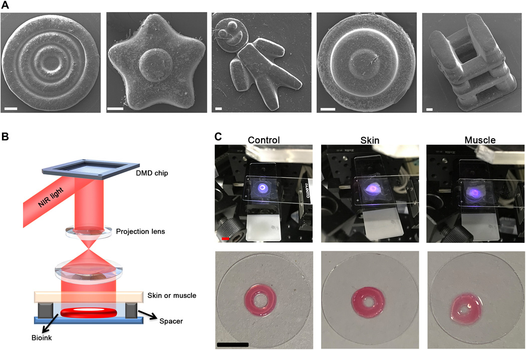
The researchers’ method involves creating a computer-aided design (CAD) model, which via a connected Digital Micromirror Device (DMD) chip, dynamically generates the digital NIR. Infrared light is then projected, to non-invasively induce the spatial polymerization of the local injected bioink, layer by layer. Commonly used biocompatible hydrogel monomers such as gelatin methacryloyl (GelMA), were demonstrated to be compatible with polymerization under this NIR irradiation process. Once the biomaterials had been activated, images were fed to the computer in sequence.
Testing showed that the DNP process was capable of quickly printing the GelMA-derived hydrogel obstacles, taking around 15 seconds to print a 200-μm-thick layer of tissue. In order to evaluate the capacity of the method, three-ring microconstructs with decreasing widths from 200 to 100 μm, were fabricated using the DNP process. Double-layered microstructures with precise, customized 3D features in flower-like shapes, cake-like shapes and a type of truss structure, were also produced ex vivo, proving the capabilities of the technique.
The researchers covered a piece of 0.5-mm-thick pig muscle tissue in the bioink, in order to mimic the phenomena of non-invasive in vivo 3D bioprinting. Using the NIR light, the team were able to excite a patterned emission from the nanoinitiators in the bioink, and induce polymerization. When the procedure was tested inside lab mice, the process did not impact on their surrounding tissues, which had complete tissue structures, without significant inflammation or abnormal defects. Three types of structures, including triangle, cross, and two-layer cake-like hydrogel constructs, were successfully non-invasively printed by the DNP process in vivo, indicating that the process is capable of non-invasive in vivo 3D bioprinting.
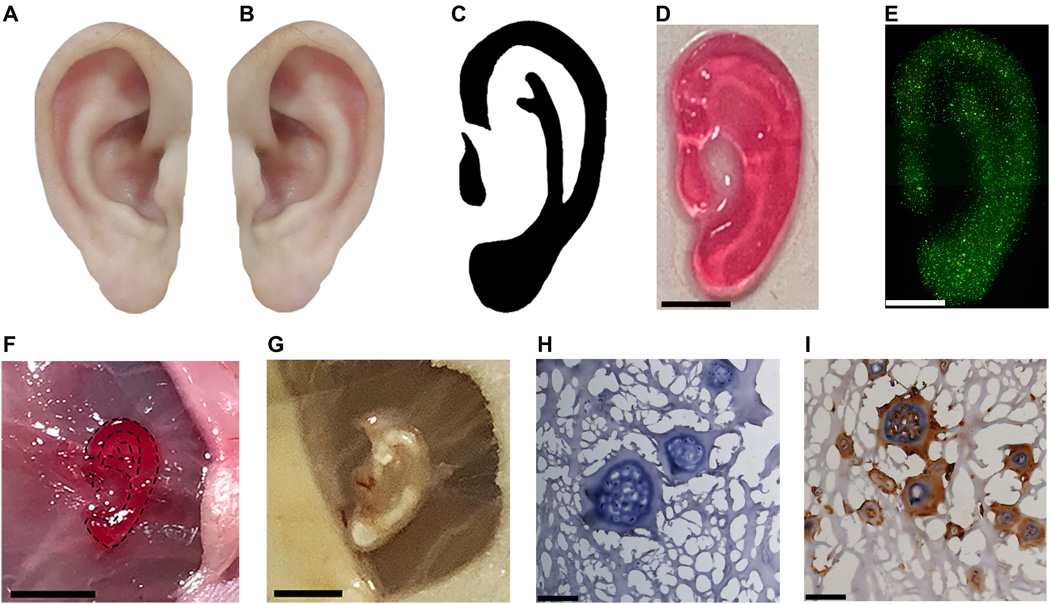
Replicating this process, the researchers created a customized ear-shaped construct containing chondrocytes. The ear’s cells had good viability after printing, cultured for 7 days in vitro, and after a month, the ear shape of the construct had been maintained. Produced as a proof of concept for non-invasive 3D bioprinting, the ear-like tissue offers promising applications in future tissue regeneration and auricle reconstruction.
Previous approaches to 3D bioprinting
Bioprinting has been used in additive manufacturing to create a range of living organs and tissues for implantation.
3D Bioprinting Solutions for instance, a Russian bio-technical research laboratory, has 3D bioprinted bone tissue in zero gravity on the International Space Station (ISS). Using the lab’s magnetic 3D bioprinter Organ.Aut, the project aims to enable the creation of bone implants for astronaut transplantation during long-term interplanetary expeditions.
Scientists from Carnegie Mellon University (CMU), Pennsylvania, used a novel 3D bioprinting method to build functional parts of the human heart, in August 2019. Using Freeform Reversible Embedding of Suspended Hydrogels (FRESH) technology the team were able to 3D print collagen for small blood vessels, valves, and beating ventricles.
3D bioprinting technology company Aspect Biosystems announced a collaboration with the Dutch Maastricht University (UM) in January 2019. The focus of the project was to develop viable kidney tissue for medical testing.
The researchers’ findings are detailed in their paper titled “Noninvasive in vivo 3D bioprinting,” published in the Science Advances journal. The study was co-authored by Yuwen Chen, Jiumeng Zhang, Xuan Liu, Shuai Wang, Jie Tao, Yulan Huang, Wenbi Wu, Yang Li, Kai Zhou, Xiawei Wei, Shaochen Chen, Xiang Li, Xuewen Xu, Ludwig Cardon, Zhiyong Qian and Maling Gou.
You can now nominate for the 2020 3D Printing Industry Awards. Cast your vote to help decide this year’s winners.
To stay up to date with the latest 3D printing news, don’t forget to subscribe to the 3D Printing Industry newsletter or follow us on Twitter or liking our page on Facebook.
Looking for a job in the additive manufacturing industry? Visit 3D Printing Jobs for a selection of roles in the industry.
Featured image shows the Noninvasive 3D bioprinting process behind the ear-shaped construct created by the researchers. Image via Science Advances.
