
Technology’s greatest accolade is perhaps its ability to facilitate connections between people in pursuit of a common goal. When applied to medicine, the implications become particularly pertinent, especially considering the treatment of fatal and almost universally felt diseases like cancer.
Digital imaging and 3D printing platforms have been developed with the expressed purpose of creating much-needed links between medical specialists and patients. At Phoenix Children’s Hospital, Arizona, such technologies have been applied to the diagnosis and treatment of a number of complicated conditions, including cancerous and defective hearts.
Getting a hold on cancer
Phoenix Children’s Hospital is equipped with a 3D Innovation Lab, giving staff access to the latest visualization and quantification technologies. The suite includes the access of IntelliSpace Portal 10 software from global technology company Philips (NYSE: PHG).
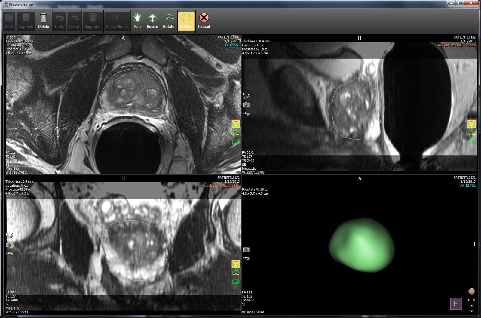
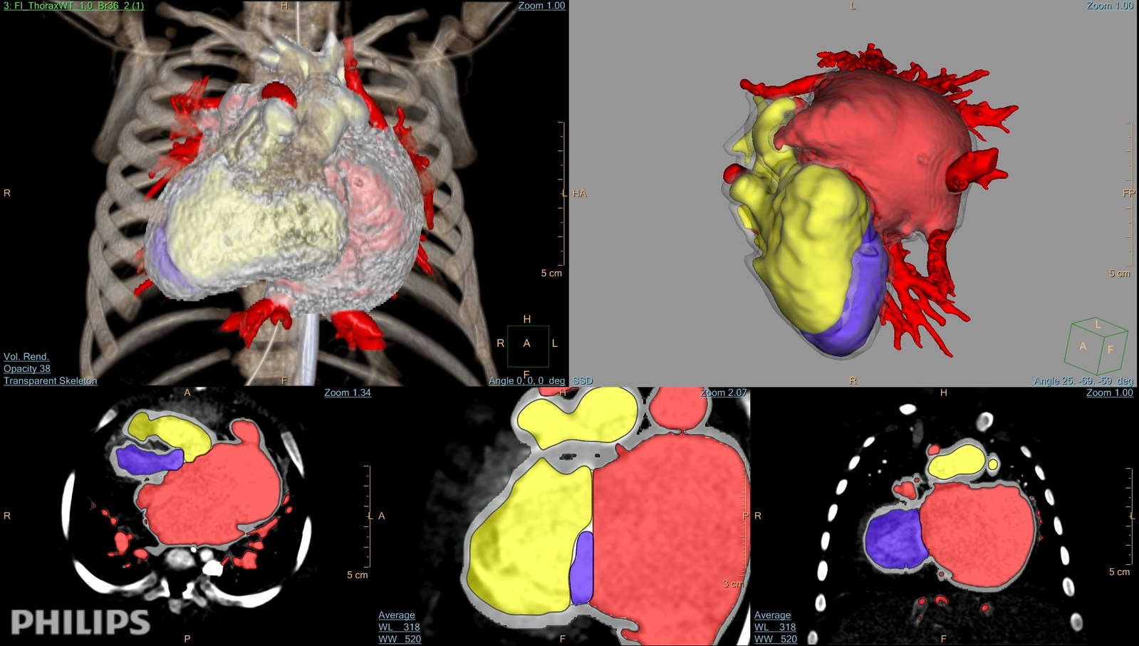
With this portal, users can create clear 3D images of a patient’s internal organs using CT and MRI scan data. Tumors can then be isolated from the images to characterize the disease and monitor its response to chemotherapy.
Dr. Richard Towbin is Chief of Radiology at Phoenix Children’s Hospital. Addressing the the hospital’s specialist 3D Innovation Lab, Dr. Towbin says, “We’ve made a big effort in the department of radiology to better present imaging data to the users of that information.”
“Any time we get digital data we can morph it into 3D, display it in 3D, and then, with the materials and unique data that we’re getting, make decisions which are going to improve the clinical outcome.”
A universal solution
With an added 3D modeling application, images in Philips software can be sent to other areas of the hospital for examination, or transformation into a physical model.
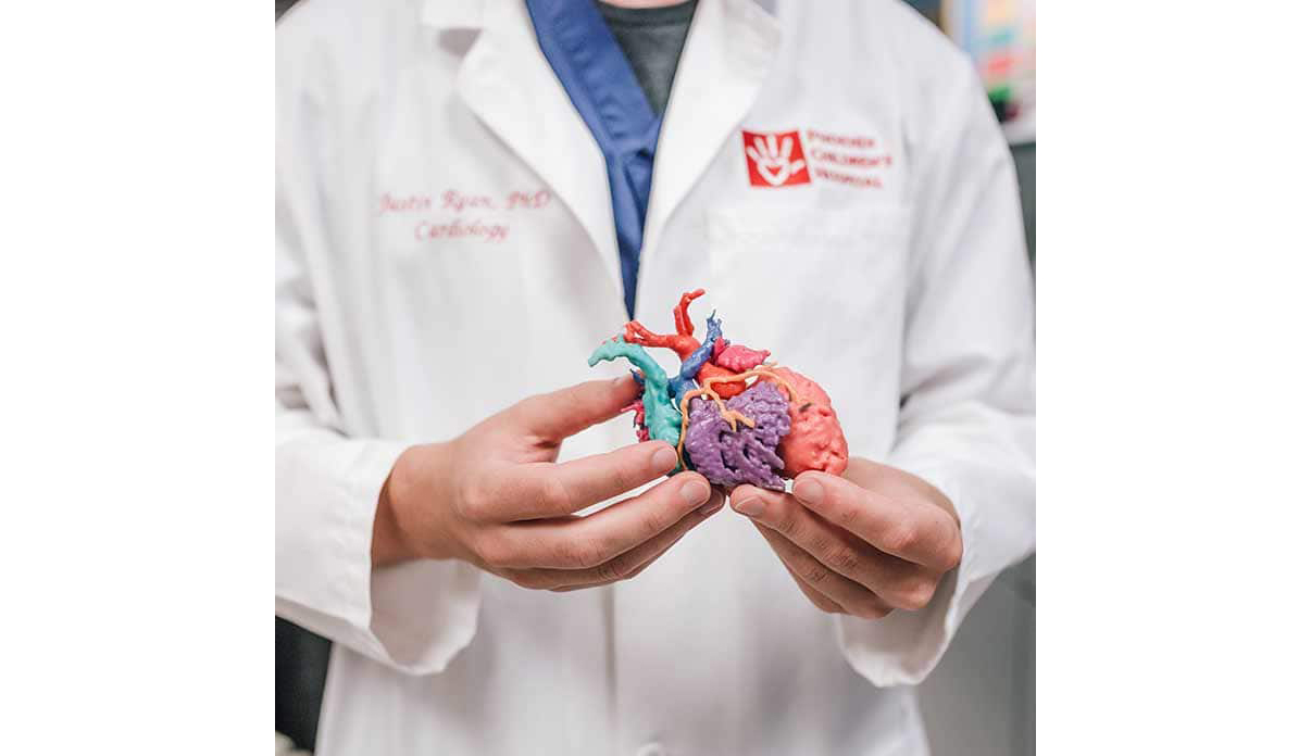
At the Cardiac 3D Print Lab, for example, surgeons use the digital imaging workflow to create test models for operations. Justin Ryan, Research Scientist at Phoenix’s Cardiac 3D Print Lab explains, “If it’s necessary for the surgeon or patient-family to better understand the anatomy, we can make a 3D printed image of the heart.”
“We can allow a doctor or a family to hold a heart in the palm of their hand.”
Dr. Dianna Bardo, Co-Director of the 3D Innovation Lab adds, “When I model a tumor or model a heart or bone into a 3-dimensional object in our 3D Lab, it’s an instant picture.”
“It’s something anybody can perceive.”
Lest wasteful, more accurate
In 3D, it is also much easier to gain an accurate perception of the actual tumor inside a patient’s body, eradicating inconsistencies that often occur when working only with a 2D image. As Dr. Bardo explains, “We’ve had 11 radiologists measure 100 tumors in 100 different patients, and everybody’s perception of the shape of the tumor varies considerably.”
This explains in part the rising number of dedicated medical model 3D printing programs, such as services from Whiteclouds and Stratasys Direct.
Phoenix Children’s Hospital’s Dr. Towbin concludes, “3D is a great example of [reducing waste and inefficiency] because now we have to do less imaging because we’ve answered questions that previously we weren’t able to answer with the 2D data.”
For all the latest news about the 3D Printing Industry, subscribe to our free newsletter and follow our active social media accounts.
Featured image shows 3D imaging to modeling in IntelliSpace Portal 10. Image via Royal Philips
