Researchers from the University of Tokyo have successfully 3D bioprinted a scaffold-free human ‘mini-liver’. According to the research team, they were able to “construct a small portion of liver tissue that could stably maintain drug, glucose, and lipid metabolism, in addition to bile acid secretion.
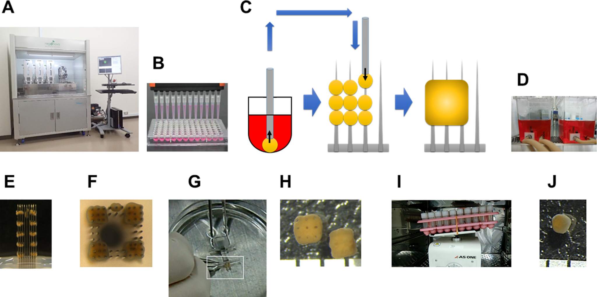
Bioprinting was performed using a Regenova bioprinter from Japanese company Cyfuse Biomedical, and this printer enables scaffold-free bioprinting with use of the Kenzan method.
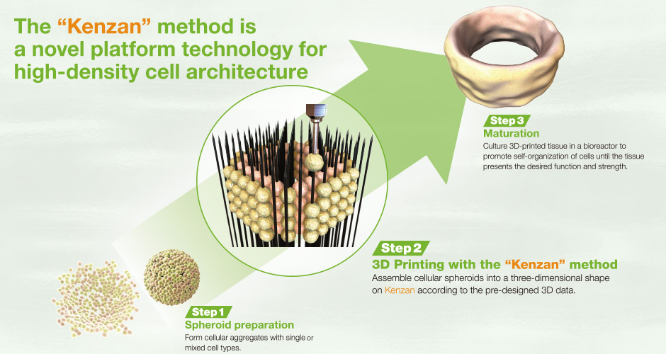
Scaffold-free bioprinting
3D printing technology has become a useful technique in the medical industry to create cell scaffolds which can facilitate the production of artificial organs. The method used by the researchers from University of Tokyo, the Kenzan approach, is significant because it removes the need for a supporting scaffold structure. It is expected that by doing so, cell tissues will have an extended functioning period. This is because they are not dependant on external support from a scaffold structure.
Kenzan method
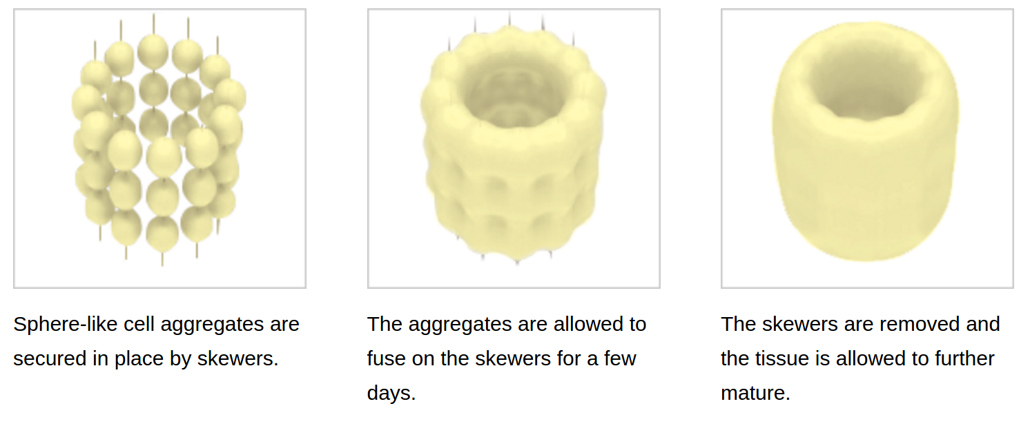
The Kenzan method was developed by prof. Koich Nakayama and Cyfuse Biomedical has exclusive rights to the technology with their Regenova printer. Using this method, the researchers secure cultured cells to needles that act as skewers. Following this, these cells are then “cultured in a perfusion chamber for 4 days.”
The cultured cells grow and merge together before being removed from the skewers and furthered matured. The Kenzan method has been used recently in separate research from Japanese Kyoto University to create cells implanted into lab rats.
Having cultured the liver tissue, it was then tested for its ability to function as a typical liver tissue. Publishing their research findings in the Biochemistry and Biophysics Reports journal, the team were pleased with these results. As they state,
These results suggested that this scaffold-free bio-printing technique performed with the Regenova and hepatocyte spheroids could produce liver structures with durable functions.
The team also noted that, “these tissues exhibited both self organization and extracellular matrix (ECM) production (e.g., collagen), which are thought to contribute, at least in part, to effective liver function.” They believe their liver model is unique for its “wide range of metabolic functions” and state that it “could maintain functional metabolism of drugs, glucose, lipids, and bile acid for long periods.”
Future application of the research
In future, this unique human liver model could be widely useful in drug discovery research applications, such as safety, pharmacokinetics, identification of new drug discovery targets.
There are several companies exploring bioprinting and Californian company Organovo has suggested recently that their bioprinted livers could be sent to the FDA for approval as early as 2019. An agreement reached this week suggests that 3D Systems will now be entering the bioprinting market, so it will be interesting to see how this technology advances.
If you haven’t already, make sure to place your votes in the 3D Printing Industry Awards.
For the latest 3D bioprinting news, sign up to our newsletter and follow us on twitter and Facebook.
Featured image shows the Kenzan method. Image via Cyfuse Biomedical.