Recently in Nanning, China, a child with craniosynostosis syndrome went under a skull reshaping and augmentation operation thanks to 3D printing technology.
The five-year old boy Xiao Qiang – whose name was changed for identity security – was born in a family in the rural area of Nanjing. As he grew up, he was increasingly troubled by his appearance. Narrow skull, convex eyeballs and a much lower IQ than his contemporaries, he was even called an alien. Things hadn’t changed until he started to vomit constantly. He was taken to hospital and was finally diagnosed as craniosynostosis.
About Craniosynostosis
Craniosynostosis is a rare skull problem that causes a baby to be born with, or develop, an abnormally shaped head. The irregular skull shape in craniosynostosis can cause persistent headaches, learning difficulties, eye problems and other symptoms. Most symptoms develop in later childhood.
Craniosynostosis was first discovered in 1851. It is said that there is one diagnosed child out of every 10,000 new born babies. The best time for treatment was before two years old. The older the child gets, the harder the operation will be, and the more unlikely the IQ will recover.
The operation
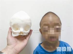
This operation was successfully accomplished by neurosurgeons from Nanjing No.2 People’s Hospital, under the lead of Professor Yao Jieming. This was the first deformity reconstructive surgery for a craniosynostosis patient with the help of 3D printing technology in the Guangxi province in China.
Neurosurgeons printed a model of Xiao Qiang’s skull and simulated the operation on the model first. The operation process may sound a little terrifying and may even remind us of a live version of The Bone Collector. The cause for the convex eyeballs is due to the undersized eye sockets. Therefore, the surgeons needed to take out the eye sockets and move their places in order to enlarge the sockets. Then the surgeons would remove the the anterior skull and divide it into six parts. With a certain rotation of each part, surgeons would put them back using some stretchable and absorbable clips so as to create some room between each part. Later on, these spaces would gradually close as the skull continues to grow.
Above is a simple explanation of how the skull is enlarged and how the pressure within the skull is reduced to a normal level.
According to the neurosurgeons, this was only the first step of the operation. After the patient is recovered, there will be another operation on the posterior skull with the same method.
We hope that young Xiao Qiang will recover soon in the future.
Feature image: nanjixiong