Conventional optics have their limits when it comes to seeing objects that are smaller than the wavelengths of visible light. The science of imaging cells generally requires chemical processing in order to make them visible under a microscope, and this treatment often kills the cells while preserving them. A company called NanoLive has developed what they call the “3D Cell Explorer”. They claim to have invented the first microscope that gives users the opportunity to see inside living cells as they are, without preparation. The process works by technology similar to MRIs, and a unique (and proprietary) software that employs holographic algorithms.
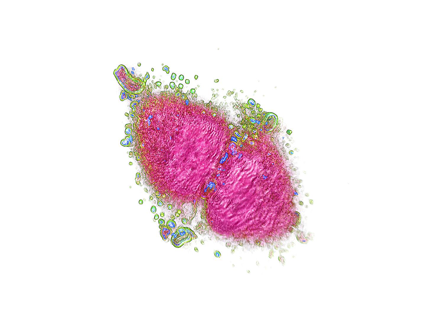
The 3D Cell Explorer is the definition of state-of-the-art and cutting edge in terms of both hardware and software. Producing unbelievably sharp 3D images of entire living cells in seconds, the 3D Cell Explorer has a higher resolution than any available conventional microscope. The device takes photographs at different depths creating “photographic slices” which are recombined using their holography software. The software digitally marks the cells, and labels the different parts. What the user is left with is “a 3D image of the cell that can be rotated and explored in depth.”
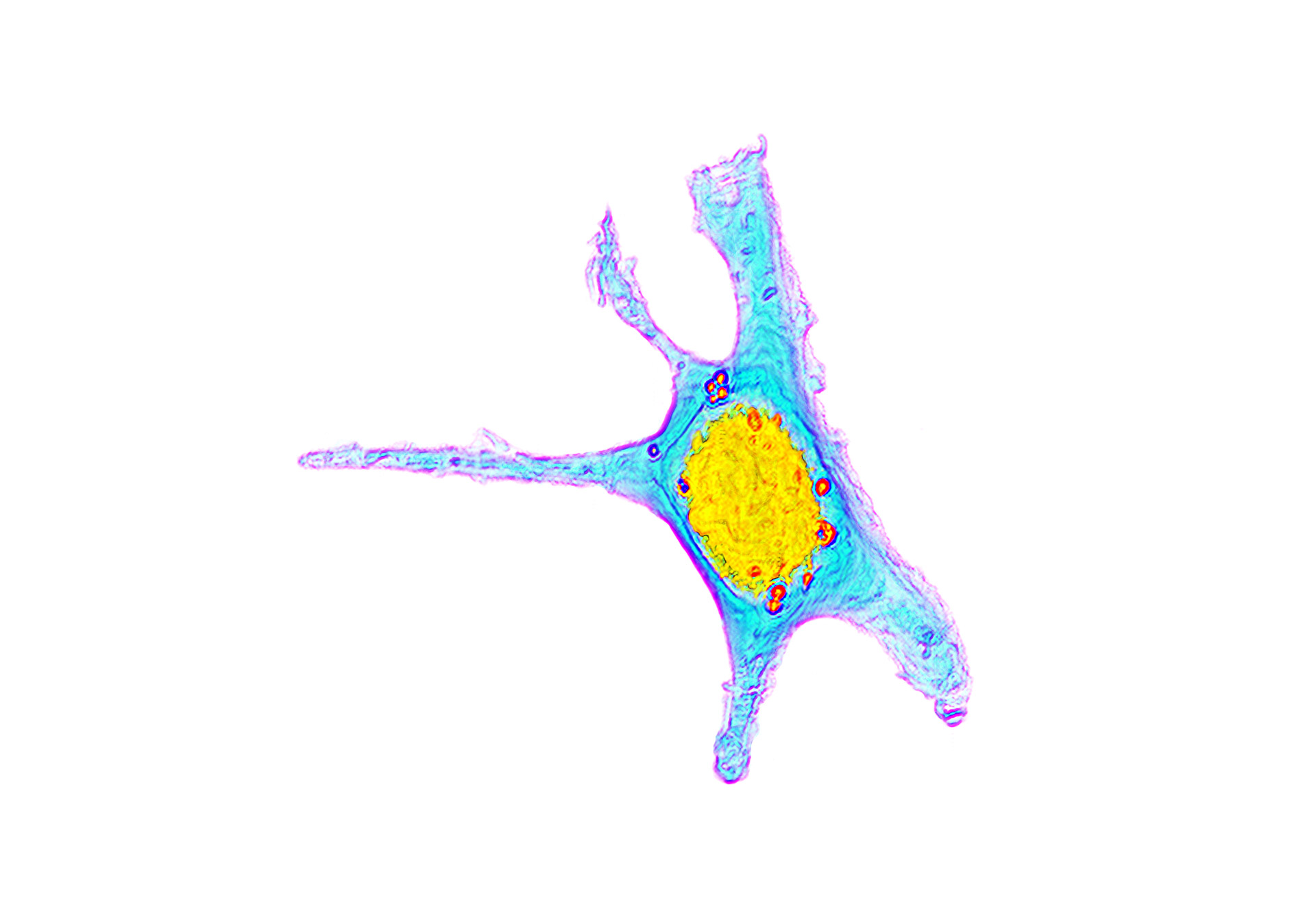
The software is hilariously named STEVE, and, as of now, is available to download. If you want to take a trip inside virtual and living cells, STEVE is for you. What allows it to “automatically define all the different parts of a cell based on an optical property” is something called the “refractive index”. Different organelles within the cell have various refractive indices, and STEVE tells them apart, and digitally marks them with either the same or different colors. Since this type of digital processing is quantitative, it can use an infinite amount of colors. This allows users to investigate changes as they occur in the cell. Real-time tracking. STEVE allows the user to constantly modify the digital markings, save them, and reuse them for different cells. As NanoLive CEO Yann Cotte puts it, “Starting from today, scientists, medics and students all around the world will be enabled to travel inside 3D cells in full color by simply downloading STEVE on their laptop.”
Nanolive’s bio is as follows: “Nanolive SA was founded in November 2013 at the EPFL Innovation Park (Lausanne) by Yann Cotte (CEO) and Fatih Toy (scientific advisor), who had just finished their PhDs at EPFL. Currently based in Ecublens, the company pioneered the 3D Cell Explorer in 2013, publishing their invention in Nature Photonics.” If you are interested, you can pre-order the 3D Cell Explorer from NanoLive. STEVE can be downloaded right here.