
California-headquartered medical imaging company QT Ultrasound has used its detailed pictures of microanatomy to 3D print a model of a woman’s breast tissue. A tangible model of the complex duct system, the model presents a potential avenue for diagnosticians working with breast cancer.
“We are on a mission to change the paradigm of breast imaging,” explains QT Ultrasound CEO Dr. John Klock.
“The benefits of being able to see breast tissue information in a true 3D volume, and then segment and isolate the tissue types, include new opportunities to examine the ductal network of the female breast in ways we never could before.”
A new paradigm in breast imaging
QT Ultrasound’s flagship product is the QTscan system. As the company name suggests, the QTscan is an imaging tool that uses ultrasound, a non-invasive method as opposed to other techniques such as an MRI. At present, the technology is not yet ready to succeed conventional mammography, however it is specifically designed for picturing the breast.The company seeks to improve the accuracy of cancer diagnoses, especially for dense breast tissue which can result in tumors being missed.
In 2018, the QTscan system was granted clearance by the FDA as an adjunct to mammography. Dr. Klock adds, “Transmission ultrasound provides high definition imaging of breast tissue, and we’re excited to show why true 3D data acquisition and image reconstruction matters.”
Detecting breast cancer with 3D prints
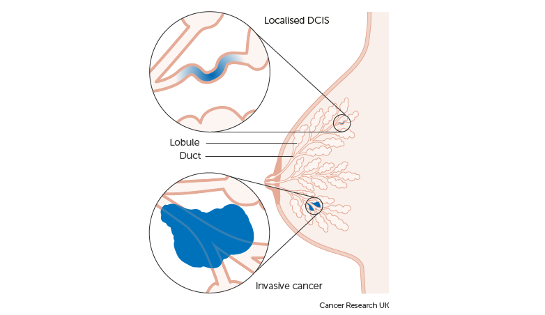
The 3D printed model from QT Ultrasound depicts the the inner network of a woman’s breast ducts. This is where the earliest form of breast cancer (Stage 0 and 1 ductal carcinoma in situ) occurs, and it is typically non invasive, as the abnormal cells have yet to spread into surrounding tissue. Though more common in women, men can also contract ductal carcinoma in situ (DCIS). In the UK, according to recent figures from Cancer Research, around 6700 people are diagnosed with DCIS each year, including about 25 men.
The key with DCIS is detecting it early, which can be achieved through regular screening and examination. QT Ultrasound’s 3D printed model could be used in a further investigation of tissue abnormalities, such as lumps or discharge. In this case, not only can it be used by physicians to better understand the condition and consider potential treatments, it can also be used as a communication aid with the patient. This very practice is being applied to the treatment of numerous medical conditions all around the globe.
In 2018 3D Systems launched a specialist On Demand Anatomical Modeling Service for the purpose, and Phoenix Children’s Hospital, Arizona is just one of the institutions with a dedicated facility for 3D printing. Dr. Dianna Bardo, Co-Director of Phoenix Children’s Hospital’s 3D Innovation Lab explains, “When I model a tumor or model a heart or bone into a 3-dimensional object in our 3D Lab, it’s an instant picture. It’s something anybody can perceive.”
For more medical 3D printing updates subscribe to our newsletter, follow us on Twitter and like us on Facebook. Visit 3D Printing Jobs for new opportunities in your area.
Featured image shows a 3D printed model of breast ducts. Clip via QT Ultrasound
