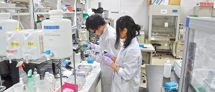
Jichi Medical University School of Medicine Department of Neurosurgery in Japan have developed a method for manufacturing solid models of cerebral aneurysms, using a compact 3D printer with ABS resin. This has been done before, but the team has shortened the printing time from previous conventional methods. With further investigation, the team managed to apply the method to emergency clipping surgery often associated with severe brain aneurysms.
What is a cerebral aneurysm?
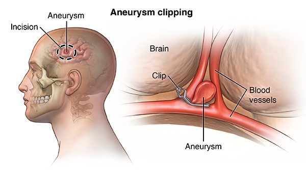
A cerebral aneurysm (also known as an intracranial or intracerebral aneurysm) is a weak or thin spot on a blood vessel in the brain that balloons out and fills with blood. The bulging aneurysm can put pressure on a nerve or surrounding brain tissue. It may also leak or rupture, spilling blood into the surrounding tissue (called a hemorrhage). Some cerebral aneurysms, particularly those that are very small, do not bleed or cause other problems. Cerebral aneurysms can occur anywhere in the brain, but most are located along a loop of arteries that run between the underside of the brain and the base of the skull (Courtesy National Institute of Neurological Disorders and Stroke).
Why do we need a 3DP fix to this?
Brain aneurysms, when ruptured, can be fatal. Statistics say that there is a rupture every 18 minutes, and in around 40% of cases it results in death. Of those who survive, about 66% suffer a permanent neurological deficit. Approximately 15% of patients with aneurysmal subarachnoid hemorrhage (SAH) die before reaching the hospital. Any solution to this is a welcome one, and there have been many advances involving 3D printing.
What are the team at Jichi University doing differently?
At the University, a total of 16 patients diagnosed with acute subarachnoid hemorrhage resulting from cerebral aneurysm rupture were used in the study of the technology, with emergency clipping being performed on the day of hospitalization. Digital Imaging and Communication in Medicine (DICOM) data obtained from computed tomography angiography (CTA) scans were edited and converted to stereolithography (STL) file formats, followed by the production of 3D models of the cerebral aneurysm using the 3D printer.
On average, the time from the moment of hospitalization to surgery was 242 minutes. With the implementation of the 3D printed model, the emergency clipping device can be placed much more quickly and efficiently. Originally timed at 67 minutes, the printing time has been reduced to approximately an hour after more experience was gained.
Although 3D printing is often considered to involve huge costs and long manufacturing time, the method used in the study required shorter time and lower costs than conventional methods for manufacturing 3D cerebral aneurysm models, thus making it suitable for use in emergency clipping.

You can read more about the University and their research projects at their website.