In 3D bioprinting for tissue regeneration, the goal is to deposit living cells into an environment as close as possible to the human body. One of the key ingredients for sustenance is protein, or a steroid hormone, allowing the cells to feed for a given amount of time.
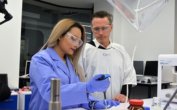
Working at Victoria’s $143.6 million Aikenhead Centre for Medical Discovery (ACMD) Swinburne PhD candidate Lilith Caballero Aguilar is looking at a way of providing protein sustenance, through the fabrication of so-called ‘microspheres’.
Food for cell growth
Proteins and hormones needed for cell proliferation are known collectively as ‘growth factors’. Potential examples of growth factors include chitosan, a protein extracted from the shells of shrimp, crabs and other crustaceans.
There are also specialist fibroblast growth factors for stimulating blood vessel differentiation, and morphogenetic proteins that encourage bone growth
To encourage single cells to grow and develop into tissues, e.g. cartilage or muscle, growth factors must be released gradually for weeks or more at a time.
Gradual release
In Augilar’s research, growth factors are encapsulated inside polymeric microspheres. The spheres are made using an emulsion of oil and water, shaken intensely, and chemically joined by crosslinking.

The spherical shape ensures material consistency and predictability, allowing the polymer to be added to 3D printer ink.
Over time, the polymer breaks down, drip-feeding cells its protein content.
Direct-to-skin 3D printing
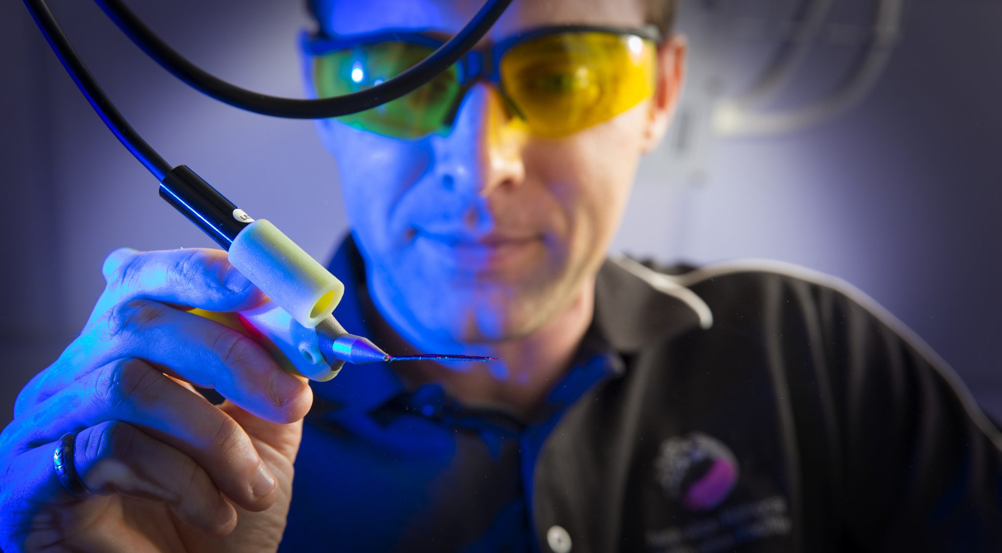
In addition to in-vitro experiments with microsphere-infused ink the BioFab3D facility at ACMD, of which Augilar is part, hopes the material can be used for direct-to-skin 3D bioprinting treatments.
To this end a group of researchers from St Vincent’s Hospital, Melbourne, and University of Wollongong are currently working on the development of a Biopen device. Speaking to 3D Printing Industry, Wollongong Professor Gordon Wallace shared that the Biopen co-axial printer has proved successful in surgical trials on sheep.
For more of the latest medical research sign up to the free 3D Printing Industry newsletter and check us out on Facebook and Twitter.
For 3D printing specific jobs in research, register on our specialist site here.
Featured image: A brain on the bench, 3D printed at BioFab3D@ACMD. Photo via BioFab3D on Twitter
