
A team of researchers at Stanford University’s School of Medicine, located in California, are developing 3D printed cardiac catheter surgical devices to help those suffering from common heart diseases, such as atrial fibrillation.
Cardiac catheter devices are used by surgeons to map a heart’s electrical activity, ultimately detecting rhythm disturbances in a patient’s heartbeats. However, these devices are often limited to one size, which results in poor connections and missed signals when surveying irregular electrical activity.
Kevin Cyr, a second-year MD student at Stanford Medicine explained that with the use of MRI or CT scans, which records an image file of a patient’s heart, the data can be fed into a 3D printer to “replicate that natural geometry and anatomy specific to that patient.”

Accuracy with every heartbeat
According to Stanford Medicine, atrial fibrillation, a heart condition causing an irregular heart rate, is the most common rhythm disorder, affecting over 6 million Americans every year. This condition disrupts the flow of blood from the heart to the rest of the body, which can lead to blood clots.
Anson Lee, Assistant Professor of Cardiothoracic Surgery at the Stanford University Medical Center began his research into common heart diseases a few years ago and was later joined by Cyr, who became interested in developing new medical technologies.
“Finding and understanding rhythm disturbances in patients has been challenging because of the one-size-fits-all nature of existing medical devices, which use electrodes that contact the surface of the heart to measure electrical activity,” explained Cyr.
Thus, the researchers leveraged additive manufacturing to create customizable cardiac-mapping catheters for each patient, which conform to the unique contours of an individual’s heart.
The 3D printed cardiac-mapping catheters
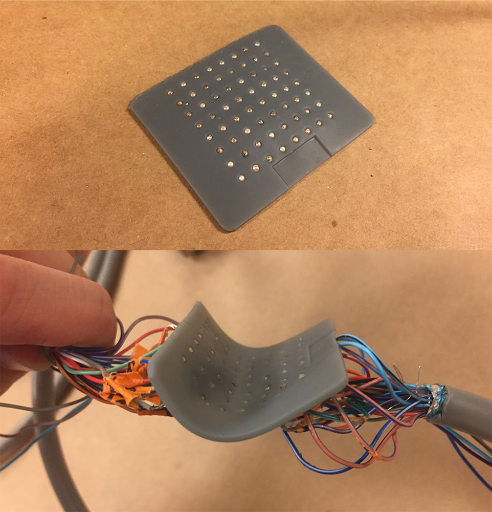
The 3D printed cardiac-mapping catheter device includes a flexible silicone membrane with small apertures in a grid-like formation which holds tiny electrodes. When placed on the surface of the heart’s atrium, the upper chamber in which blood enters the heart, the device surveys the electrical activity over that specific region.
The data is then transmitted to a computer, where it generates a recording of that heartbeat activity. The recordings produce an accurate heatmap of the electrical activity that physicians use to pinpoint the heart regions in need of treatment. Cyr added:
“We can map in perfect detail this rectangular grid of information and not have to worry about missing signals, poor contact or things like that, which otherwise might throw out errors.”
Although this device is only applicable to the external surfaces of the heart, the Stanford research team are investigating whether their device could be used to map the interior surface of the heart – further increasing the measurement accuracy of rhythmic disturbance.
Cyr believes that the devices will likely take another year or two to develop before it is tested on humans.

3D printing and cardiology
Earlier this year, researchers at the Great Ormond Street Hospital (GOSH) in London began developing 3D printed replicas of children’s hearts to be used within the pre-planning surgical process.
Prior to this, the Children’s Heart Research and Outcomes Center (HeRO) developed 3D bioprinted valves, leaflets, and patches to provide a long-lasting solution for children born with heart conditions such as congenital heart disease.
Never miss out on the latest news in 3D printing by subscribing to the 3D Printing Industry newsletter. Also, follow us on Twitter, and like us on Facebook.
Seeking new talent? Or looking for a new position? Search and post 3D Printing Jobs for opportunities and new talent across engineering, marketing, sales and more.
Featured image shows the 3D printed cardiac-mapping catheter devices. Photo via Kevin Cyr.