

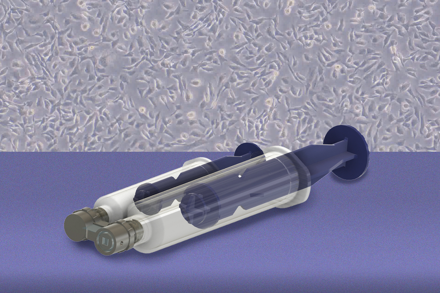
Researchers at the Massachusetts Institute of Technology (MIT) and Johns Hopkins University have developed a handheld tool built around a 3D printed microfluidic device. It collects living cells from selected spots on freshly excised tissue and leaves the rest of the tissue available for pathology. The team says the method could support earlier detection of ovarian cancer and many other cancer types, and could eventually be used to test how an individual patient’s cells respond to treatment. The findings appear in the journal Device.
Ovarian cancer is among the deadliest forms of the disease, in large part because it is often diagnosed late. Many cases begin in tissue that is difficult to sample at an early stage.
Where the Disease Starts and How Tissue Is Processed
Five-year survival for ovarian cancer can exceed 90 percent when the disease is found early, and it drops to less than half that figure at stage 3 or 4. Research from about two decades ago showed that many cases of high-grade serous ovarian cancer, the most common type, start in the fallopian tubes, where precursor lesions can be microscopic.
In the standard workflow, surgically removed fallopian tubes are placed in a chemical preservative and cut into sections. A pathologist then examines them under a microscope for cancerous or precancerous areas. The preservative keeps the tissue structure intact, but the cells can no longer be grown in culture. “It’s very time-consuming and destructive to the cells,” said Kripa Varanasi, Maher A. Elmasri Professor of Mechanical Engineering at MIT and senior author of the study.
The link between fallopian tubes and ovarian cancer has also given women at elevated risk a preventive option: removing the tubes once they no longer plan to have children, which largely preserves normal hormone production. Rebecca Stone, a gynecologic oncologist at Johns Hopkins University School of Medicine, has long advocated for the procedure in certain high-risk patients.
Angela Belcher, a professor of biological engineering and materials science at MIT, introduced Stone to Varanasi. The group then secured funding from Break Through Cancer, a foundation that assembles interdisciplinary teams around difficult cancer problems. The cell-collection tool is one component of their broader early detection project.
Fluid Flow in Place of a Scalpel
The concept took shape after the MIT team observed surgeries at Johns Hopkins. Varanasi’s lab studies how fluids interact with surfaces, and surgeons had mentioned that cells in some regions came loose during washing. “We realized we could use a fluid instead of a scalpel or brush, because when you flow a fluid it applies shear stress at the interface,” Varanasi said.
The resulting device is 3D printed and works without chemicals or enzymes. “We came up with this device where one syringe creates a vacuum that holds it against the tissue, and a second syringe pushes liquid through it,” said Bert Vandereydt, an MIT postdoc and co-first author. “The vacuum creates a seal, so nothing leaks, and then we locally apply what is basically a microfluidic chip on the tissue that gently shears the cells off.”
Testing on Human Tissue
The researchers showed that the force applied to tissue could be adjusted, and they benchmarked the method against other cell collection workflows. Cells harvested with the device stayed viable and grew in culture more readily than cells detached by conventional techniques.
The team then worked with fresh human fallopian tubes shipped from Johns Hopkins, often on short notice. After the experiments, the samples went back for conventional pathology. “We were able to collect living cells from those fallopian tubes and turn those into organoids, which is important for testing, disease modeling, and eventually developing personalized treatments,” Vandereydt said.
Different cell types required different shear levels. “There are very loosely adherent prostate cancer cells that detach at 1 pascal [of stress], but if you look at bone cancer cells, only a few cells detach under as high as 5 pascals of stress,” Vandereydt said.
Belcher said her group is working on optical methods to flag suspicious tissue regions. The device could then retrieve living cells from those specific sites for further analysis.
Path Toward Clinical Use
Early applications will focus on tissue already removed from the body, which the researchers see as a simpler route to regulatory approval. Longer term, they want the device to sample tissue inside patients.
Stone said the tool could eventually be built into standard histopathology workflows. That would give researchers a way to study how cancer forms and to investigate basic biology directly from human tissue.
According to Varanasi, researchers in the Break Through Cancer network working on pancreatic cancer, brain cancer, and leukemia pointed to a common need. “What we heard again and again is how valuable it would be to have better ways to obtain living cells from specific regions of tissue,” he said.

3D Printed Microfluidics and Living Cells
The MIT and Johns Hopkins tool depends on a 3D printed microfluidic device that moves liquid across tissue without killing the cells it collects. The cells have to survive contact with the printed part for the approach to work.
Researchers at Queensland University of Technology (QUT) have tested whether 3D printed microfluidic parts can support cell-based work. They printed culture channels, pillar arrays and droplet-generator channels on Asiga UV Max X27 DLP printers, using two resins from DMG Digital Enterprises. Liver cells from the HepG2 line were seeded into the printed channels and showed limited cell death after five days, and the team concluded both resins were biocompatible. The QUT work connects to the MIT device through viability: living cells in contact with a 3D printed microfluidic part. It differs in use. QUT grew a cell line inside the device, while the MIT device detaches cells from patient tissue and carries them out for culture.
Viability inside printed microfluidic parts is what lets these devices handle living samples, and the MIT study applies that to cells taken directly from human tissue.
Titled “A fluidic-shear device for minimally disruptive, site-selective collection of viable cells from excised fallopian-tube tissue,” the study was conducted by Domitille Avalle, Bert Vandereydt, Sean Parks, Huaiyao Peng, Rebecca Stone, Angela Belcher, and Kripa Varanasi.
Register now for AMA: Software 2026. Join 3D Printing Industry on 22 October for expert presentations, panel discussions and live audience Q&A exploring the software shaping additive manufacturing. Register here.
To stay up to date with the latest 3D printing news, don’t forget to subscribe to the 3D Printing Industry newsletter or follow us on LinkedIn.
Explore the full Future of 3D Printing and Executive Survey series from 3D Printing Industry, featuring perspectives from CEOs, engineers, and industry leaders on the industrialization of additive manufacturing, 3D printing industry trends 2026, qualification, supply chains, and additive manufacturing industry analysis.
Featured image shows MIT Ovarian Detection. Image via MIT.