Researchers from Boston University College of Engineering have developed a new method for treating ischemia, by 3D printing a vascular patch which encourages blood vessel growth.
Ischemia relates to inadequate blood supply to an organ, in this case the heart, and as the research cites in the paper – ischaemic heart disease is the leading cause of death in the U.S and continues to become more prominent. Currently, treatments such as surgery are generally limited to treating large arteries. While the new method hopes to provide a solution for the microvasculature level.
While 3D printing has been explored before to create blood vessels, this research team led by Christopher Chen is keen to emphasis the importance of structural growth. When creating artificial blood vessels it is important to provide organized growth in order to provide a functional solution.
Disorganized blood vessels
As Christopher Chen explains, there are other experimental techniques to treat ischemia such as therapeutic angiogenesis which involves injecting “growth factors” to encourage vessel growth. However according to Chen, “the new branches that sprout form a disorganized and tortuous network that looks like sort of a hairball and doesn’t allow blood to flow efficiently through it. We wanted to see if we could solve this problem by organizing them.”
The research team then tested this theory by implanting organized and disorganized structures into rodents. Bioprinted blood vessels have been tested on mice before with research from University of California, San Diego that was able to transport nutrients around the body. Chen explains the importance of this part of the research as,
One of the questions we were trying to answer is whether or not architecture of the implant mattered, and this showed us that yes, it does, which is why our unique approach using a 3D printer was important,
Research method
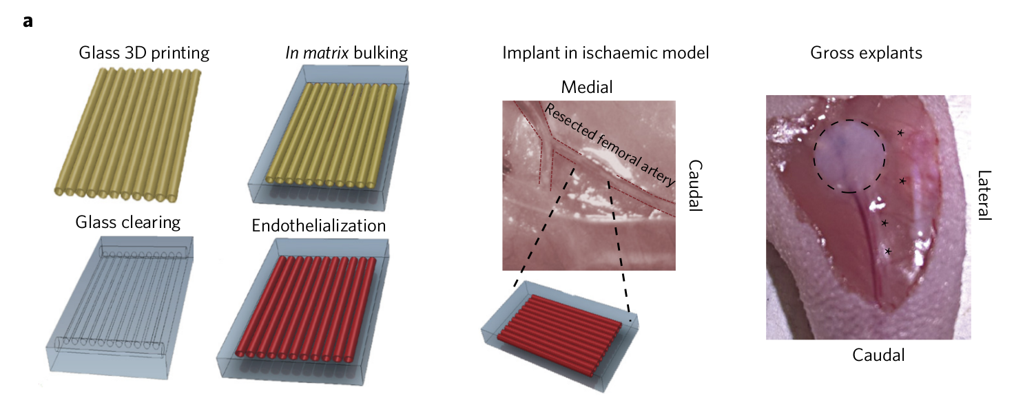
Chen’s team turned to bioprinting through local Boston company Innolign and the paper explains what this process entailed,
To engineer endothelial networks, we used a recently described method to 3D print sugar filaments into a network, encapsulate the network in a fibrin gel, dissolve the sugar to leave behind channels and inject endothelial cells to fill the channels.
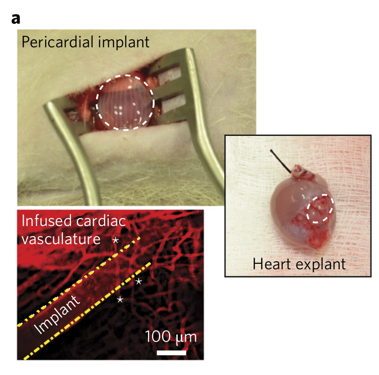
Chen reports the success of this process as the bioprinted patch “helped to guide the formation of new blood vessels that seemed to deliver sufficient blood to the downstream tissue. While it wasn’t a full recovery, we observed functional recovery of function in the ischemic tissue.” In a similar way, research from Oregon Health & Science University enlisted bioprinting to create molds for blood vessels inside teeth as an alternative root canal treatment.
Following this initial success, Chen and his team will work on scaling the process and further refining the method. The paper, titled ‘3D-printed vascular networks direct therapeutic angiogenesis in ischaemia’, has been published in Nature Biomedical Engineering.
For all the latest bioprinting news, subscribe to the most widely read newsletter in the 3D printing industry, follow us on twitter and like us on Facebook.
Featured image shows the blood vessel network of the heart. Photo via Rob Jones on Flickr.