
A group of researchers in China has constructed ears for children suffering from microtia, a congenital condition where the external ear (pinna) is underdeveloped, with the help of 3D scanning and 3D printing.
The scientists, from Shanghai Jiao Tong University, the National Tissue Engineering Research Center of China, the Chinese Academy of Medical Science, Wei Fang Medical College and Dalian University, engineered a patient-specific ear-shaped cartilage in vitro using a 3D printed biodegradable scaffold and Microtia Chondrocyte (MCs) cartilage cells.
Microtia and 3D printing
Microtia can have a negative impact on the hearing and wellbeing of children affected by it. Established procedures to treat microtia include rib cartilage reconstruction, plastic implants or prostheses.
Projects like Australia’s FutureHear are 3D printing customized ear molds, while Dr. Ken Stewart of the Royal Hospital for Sick Children in Edinburgh has used 3D scanning and 3D printed models to prepare cartilage reconstructions accurately in the shape of an ear.
This approach, however, combined 3D printing with in vitro tissue engineering on children suffering from microtia only in one ear.
3D printing a guide for a healthy ear
To begin with, a scan was taken of the healthy ear and a digital image of it created using 3DPRO software. This digital image was then symmetrically mirrored to guide reconstruction.
A corresponding resin model of this mirrored ear was 3D printed on a Z Corp Spectrum 510 3D printer, a model that first shipped in 2005 by the company who would later be acquired by 3D Systems in 2012.
The 3D printed model was used to cast a pair of molds from clay and silicone, which could then hold biomaterial scaffolds.
Growing a healthy ear on 3D printed scaffolds
To produce the biodegradable biomaterial scaffolds, a 9 square centimeter inner PCL mesh with three square milimeter grids and a thickness of 1.37 mm was 3D printed.
This inner core was wrapped with PGA unwoven fibers and coated with PLA. Expanded MCs harvested from the child’s microtic ear. These were evenly dropped onto the PGA/PLA layer of the ear-shaped scaffold and then placed in a cell culture solution. The cartilage ear was removed after twelve weeks.
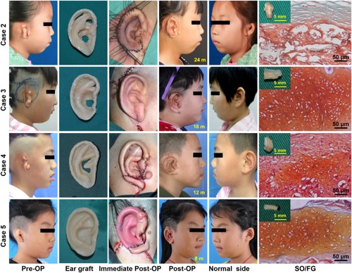
The cartilage ear was surgically implanted in a similar way to rib cartilage. The child’s skin was draped over the cartilage and it fit the new shape of the ear thanks to vacuum drainage.
Conclusions from the research
While four of the five repetitions of this study were an immediate success, the authors admit that closer analysis and process refinement may help to introduce this treatment clinically.
The full paper, “In Vitro Regeneration of Patient-specific Ear-shaped Cartilage and Its First Clinical Application for Auricular Reconstruction” by Guangdong Zhou, Haiyue Jiang, Zongqi Yin, Yu Liu, Qingguo Zhang, Chen Zhang, Bo Pan, Jiayu Zhou, Xu Zhou, Hengyun Sun, Dan Li, Aijuan He, Zhiyong Zhang, Wenjie Zhang, Wei Liu, and Yilin Cao is available to read online.
We want to know about the most important advances in additive manufacturing. Make your nominations for the 3D Printing Industry Awards 2018 now.
For more stories on 3D printing in biomedical research, subscribe to our free 3D Printing Industry newsletter, follow us on Twitter, and like us on Facebook.
Featured image shows a successfully regenerated ear-shaped cartilage. Photo via EBiomedicine.