
New research is using 3D printing to advance understanding and treatment of abnormal heart rhythms.
The heart is the most important organ in the human body. Understanding how the heart behaves is crucial for doctors conducting surgical procedures, but also of vital importance within the field of medical research.
Scientists at universities in the UK and Denmark have developed a new method of conceptualising the cardiac conduction system (CCS) – the system responsible for the heart’s automatic rhythmic beat. This new technique will mean surgeons will be able to plan complex heart surgery in abnormal hearts without damaging precious tissue.
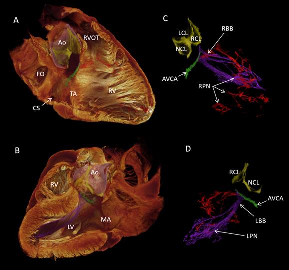
The CCS of the heart consists of several components, each of which are critical to the functioning of the heart. These components include: the Sinoatrial Node (responsible for excitation signals), Atrioventricular Node (responsible for delaying the signal), bundle of His (which transmits impulses from the Atrioventricular Node), and the Purkinje Fibres (working with the bundle of His to spread the waves along the ventricles, causing them to contract).
Lowering the risk of repairing hearts
Scientists from Liverpool John Moores University (LJMU), The University of Manchester, Newcastle University, and Aarhus University in Denmark are now able to visualise this conducting system in a 3D format via their new study. It has been titled: “High resolution 3-Dimensional imaging of the human CCS from microanatomy to mathematical modelling”. The findings were published in the academic journal Scientific Reports.
Data from the study provides a more detailed approach to analysing the heartbeat than that which is currently available on computer models. By revealing the exact location of the CCS in a normal heart, our understanding of widespread heart rhythms problems like heart block and atrial fibrillation – affecting more than 33 million people worldwide – should hopefully be improved.
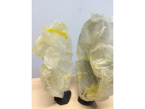
Professor Jonathan Jarvis of LJMU’s School of Sport and Exercise Sciences explained, “The 3D data makes it much easier to understand the complex relationships between the cardiac conduction system and the rest of the heart.” Once the data is captured, it can be formatted for further use. “We also use the data to make 3D printed models that are really useful in our discussions with heart doctors, other researchers and patients with heart problems,” said Professor Jarvis.
“New strategies to repair or replace the aortic valve must therefore make sure that they do not damage or compress this precious tissue. In future work we will be able to see where the cardiac conduction system runs in hearts that have not formed properly. This will help surgeons who repair such hearts to design operations that have the least risk of damaging the cardiac conduction system.”
Advancing 3D scanning for medical imaging
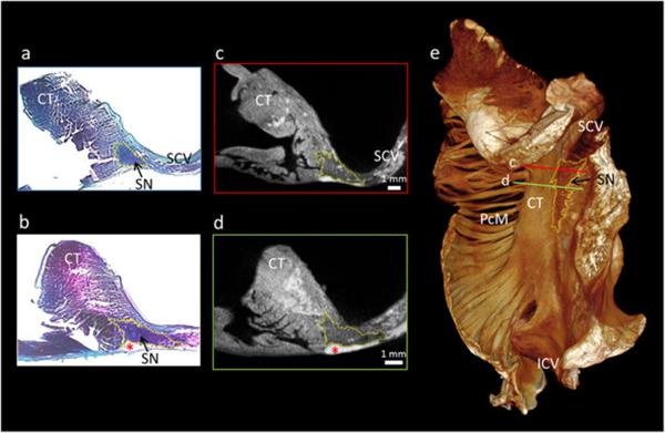
The process works by soaking post-mortem samples in an iodine solution, which means that the soft tissues can absorb X-Rays and therefore become visible. These X-Rays are then used to make 3D scans detailed enough to distinguish the boundaries between single heart cells and even detect the direction they are arranged. Micro-CT scanning was conducted using the Nikon Metris XTEK 320 kV Custom Bay and Nikon XTEK XTH 225 kV systems at the Manchester X-Ray Imaging Facility, University of Manchester.
Over the last decade, there have been a number of advancements made in understanding of the CCS, but due to its unique embryological origin, identifying the CCS through gross specimen and surgery is difficult. However, thanks to this new 3D method, anatomists and cardiovascular specialists may now be able to successfully locate the CCS by using a 3D model of its arteries.
Dr. Halina Dobrzynski of The University of Manchester’s Cardiovascular Division, who has been working on the anatomy of the CCS for 20 years, says “This is just the beginning. The British Heart Foundation is supporting my group to visualise this system in 3D from aged and failing hearts. With my research assistant Andrew Atkinson and working with Professor Jonathan Jarvis, Robert Stephenson and others, we will produce families of data from aged and failing hearts in 3D”.
For all the latest news and information about medical and healthcare applications of 3D printing, subscribe to our newsletter.
You can also follow the most popular 3D printing social media channels on Twitter and Facebook.