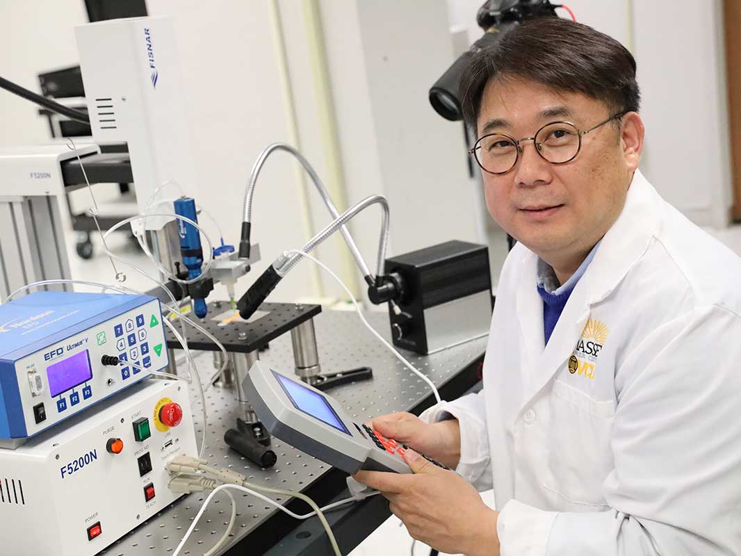
An assistant professor from the Virginia Commonwealth University (VCU) College of Humanities and Sciences has used 3D printing to create live models of tumor cells, which could enable cancer researchers to better understand the disease’s progression.
Assistant professor Daeha Joung worked alongside a team of researchers led by Dr. Michael McAlpine at the University of Minnesota (UMN), that invented the novel method of building tumor constructs. The new technique allows for the precise placement and control of living cells, to more effectively mimic the key steps of cancer dissemination. Now at VCU, Joung is using this approach to enable the university’s Massey Cancer Center investigators, to maximize cancer cell growth for study, and evaluate the effectiveness of anticancer immunotoxins.
Evolving from 2D printing in cancer research
Researchers from The European Molecular Biology Laboratory in Germany found in their study in December 2018, that 2D structures have difficulty in accurately replicating the characteristics of native tumor microenvironments. 3D cell cultures were found to establish cell-to-cell interactions that could better mimic the specificity of real tissues, doing so at greater speed, and displaying more complex behaviors.
However, according to the UMN researchers, the metastatic nature of cancer remains a major prognostic and therapeutic challenge for current models, because the movements of tumor cells are regulated by various chemical signals. A more precise model is, therefore, necessary, that incorporates these elements of tumor microenvironments, and enables the study of multilevel interactions that occur at distant parts of the body. “A number of strategies have been developed to overcome these obstacles but have not been entirely successful,” explains Joung. “I can use physics, nanotechnology, and engineering to overcome those obstacles, and provide a unique insight that could open the door to new therapeutic options for cancer.”
The team’s new approach addresses these limitations by using 3D bioprinting and an origami-inspired self-folding technique to create complex multi-material, multi-scale, and multi-functional 3D structures. These allow for the integration of numerous combinations of living cells and supporting matrices with precise spatial control, resulting in the engineering of tissues with promising biomedical applications.
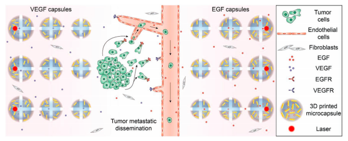
Manufacturing 3D printed cell control capsules
3D printed stimuli-responsive capsules enabled the researchers to control the cells within these structures. Containing an aqueous core, and a payload of functional molecular factors and plasmonic gold nanorods (AuNRs), the microscopic pods allow for the triggered release of chemical cues in 3D matrices. Once activated using laser radiation, the research team were able to manipulate cellular behaviors at a local level. When this was combined with 3D cellular printing, it allowed for the creation of complex tumor constructs that were controlled using multiplexed chemical signals.
To demonstrate spatial control with the capsules, the researchers printed an array of cores containing varying volumes which could be precisely triggered by deploying a UV laser. Using 3D printing enabled the researchers to test five groups of migration samples in parallel under identical conditions. This approach also allowed him to add materials where necessary, and to customize the cells produced, to show different behaviors.
As a result of the technique, Massey scientists will be able to provide cancer tissue samples to Joung, and then prescribe specific designs or tumor structures they need to have manufactured. Using robotic mechanisms and customized 3D printing technology, Joung will then adjust the amount and specific configuration of the types of cells being replicated and printed.
This, in turn, could enable future researchers to screen novel anticancer drugs, and test patient-specific strategies for diagnosis and therapeutics. “Cancer is cancer from an engineering point of view, so my lab can produce many different types of tumor tissue-mimics,” said Joung. “If someone is interested in lung cancer, we can print lung cancer cells. If someone is interested in breast cancer cells, we can print those too,” he added.
Joung’s ambition is to design a comprehensive platform of advanced 3D systems for application in cancer neural regeneration models, opening novel opportunities to test therapeutic options to treat disease.
3D printing methods of combating cancer
Scientists and researchers from other academic institutions have also used 3D printing to create live 3D cell models, with the aim of better understanding and treating cancerous tumors.
In April 2020 researchers from the USA and Germany presented a new way of studying glioblastoma (GBM), an aggressive type of brain cancer. They used a collection of human brain cells and biomaterials featuring perfused vascular channels, to allow long-term culture and drug delivery. 3D imaging technology was summarily used for the noninvasive assessment of the tissue constructs.
A master’s student from the University of Waikato, New Zealand, announced in August 2018, that she planned to grow and test cancer tumors using real cells, following the design of her 3D printed plastic tumor models. The basis for these bioprinted models was a series of webbed, palm-sized hemispheres that resembled breast cancer tumors.
Researchers from Katholieke Universiteit Leuven in Belgium, and the Indian Institute of Technology Hyderabad in India, released studies in June 2017 investigating how 3D printing can be used for cancer research. While both papers highlighted the potential of using 3D printing to create microenvironments, the Indian researchers raised the possibility of being able to precisely place cells and reproduce in vivo tumor microenvironments.
The researchers’ findings are detailed in their paper titled “3D Bioprinted In Vitro Metastatic Models via Reconstruction of Tumor Microenvironments” which was published in the Advanced Materials journal and was co-authored by Fanben Meng, Carolyn M. Meyer, Daeha Joung, Daniel A. Vallera, Michael C. McAlpine and Angela Panoskaltsis‐Mortari.
The nominations for the 2020 3D Printing Industry Awards are now open. Who do you think should make the shortlists for this year’s show? Have your say now.
Subscribe to the 3D Printing Industry newsletter for the latest news in additive manufacturing. You can also stay connected by following us on Twitter and liking us on Facebook.
Looking for a career in additive manufacturing? Visit 3D Printing Jobs for a selection of roles in the industry.
Featured image shows assistant professor Joung, who has developed a new technique for creating 3D models of tumors using live cells. Photo via VCU.
