Researchers from Rice University have developed a new method of using 3D printing to create artificial vascular networks from powdered sugar.
Replacing traditional production methods with Selective Laser Sintering (SLS) 3D printing, the team created sacrificial templates made from laser-sintered carbohydrate powders. These sugar-based constructs enable cell-laden hydrogels to be patterned with dendritic vessel networks, without the use of support materials. The newly-devised technique could improve the speed and scale of biomaterial production.
“One of the biggest hurdles to engineering clinically relevant tissues is packing a large tissue structure with hundreds of millions of living cells,” said Ian Kinstlinger, lead author and graduate student at Rice’s Brown School of Engineering. “Selective Laser Sintering gives us far more control in all three dimensions, allowing us to easily access complex topologies while still preserving the utility of the sugar material.”
“A major benefit of this approach is the speed at which we can generate each tissue structure. We can create some of the largest tissue models yet demonstrated in under five minutes.”
Devising a new and improved 3D printing method
Metabolic function in human tissues is sustained by the delivery of oxygen and nutrients, as well as the removal of waste, through complex 3D networks of blood vessels. Understanding vascular systems was essential for scientists in creating multicellular organisms, and reproducing them is equally vital to enabling 3D printed tissues to be developed. According to the research team, these tissues need to be supported using biocompatible matrices, in order to survive and supply the required nutrients to the tissue’s host.
Previous studies have seen soft lithography and needle moulding methods used to create these matrices, but 3D printing advances have led to the increased adoption of methods such as direct extrusion and inkjet-created polymers. In other research, light has been harnessed to generate sophisticated microchannel architectures, but ‘sacrificial templating’ has emerged as the dominant and most widely-used method.
The templating technique involves creating a temporary sacrificial gelatin in the shape of the desired vascular network, in which cells are encased, and then selectively removed. While extrusion-based 3D printing techniques have led to an increased adoption of this method, the features and complexity of the sacrificially templated networks have remained limited. “There are certain architectures, such as overhanging structures, branched networks and multivascular networks, which you really can’t do well with extrusion printing,” explained Jordan Miller, co-author of the study, and Assistant Professor of Bioengineering at Rice.
As a result, vascular systems printed using extrusion methods, are often subject to deformation or collapse under their own weight, and their viscosity and surface tension make precise dispensing of small volumes difficult. Moreover, printing the cells with support materials may mitigate these issues, but at the expense of longer print times and additional post-processing steps, which become increasingly difficult with increasing vascular complexity.
SLS 3D printing meanwhile, utilizes a fully supported and powder-based build volume, which enables the fabrication of objects with complicated overhangs and unsupported geometries. “Selective laser sintering gives us far more control in all three dimensions, allowing us to easily access complex topologies while still preserving the utility of the sugar material,” added Miller.
The research team hypothesized that using SLS 3D printing to produce the sacrificial materials instead of extrusion techniques, could allow vascular networks in hydrogels to be readily patterned in the presence of fragile human cells. By creating extensively branched carbohydrate filament networks via SLS, and applying them sacrificially to pattern volumetric vascular networks, the team aimed to create a faster and more stable bioprinting process.

Creating the sugar-based vascular networks
Isomalt, a sugar-alcohol commonly used in sugar-free lozenges, was found to be compatible with SLS, and the team devised a workflow for automated fabrication of 3D structures from isomalt powder. While it would have been possible to sinter a single layer of pure isomalt, the powder’s strong cohesion and relatively poor flowability made it badly suited for spreading into smooth, thin layers as required. Further mixing the powder with cornstarch was found to effectively augment the powder’s flow while preserving sintering quality. Using this concoction, the research team successfully fabricated structures with 3D branching and unsupported geometry.
Beginning with the patterning of a simple branched architecture, the researchers went on to cast a series of elastomers, stiff plastics and hydrogels around post-processed carbohydrates. During the process, the hydrogel became semi-solid within minutes, and the original template was then sacrificed, being dissolved in water or phosphate buffered saline (PBS). In each case, perfusion through the patterned channel network demonstrated channel patency and the connectivity of branched filaments. Despite the opacity of the sintered carbohydrates, polyethylene glycol and diacrylate gels were successfully polymerized by incident light from various angles, demonstrating the team’s methodology.
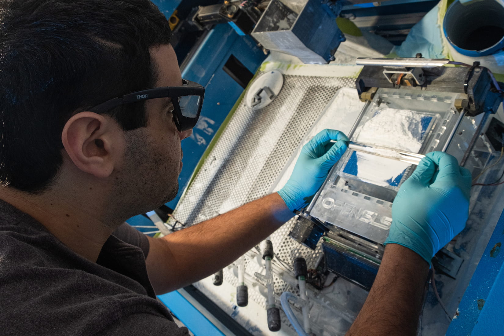
Moreover, the researchers’ newly-developed iteration of OpenSLS hardware and firmware, optimized the process for carbohydrate SLS, and an updated software toolchain was used to prepare 3D models. By manipulating the multi-extruder capabilities of an open-source slicing software designed for extrusion 3D printers, the team were able to encode specific sintering parameters, enabling them to fine tune the model’s final geometry.
Working with scientists from The University of Washington whose research group specializes in studying the delicate cells, the team later demonstrated the seeding of endothelial cells in rodent liver cells called hepatocytes. “We showed that perfusion through 3D vascular networks allows us to sustain these large liverlike tissues,” said Miller. “While there are still long-standing challenges associated with maintaining hepatocyte function, the ability to both generate large volumes of tissue and sustain the cells in those volumes for sufficient time to assess their function is an exciting step forward.”
The Rice team’s new methodology enabled them to overcome the drawbacks of previous 3D printing techniques, and produce elaborate fluidic networks within engineered living tissues. Moreover, while the researchers’ OpenSLS technique allowed them to effectively create the carbohydrates with diameters as small as 300μm, the higher quality optical components in commercial SLS printers could yet yield higher resolution templates. This opens the opportunity for further upgrades for the process. Nonetheless, the rapid nature of the manufacturing process, with none of the experiments taking longer than 15 minutes, could yet enable the process to be utilized in a range of bioprinting applications.
“This method could be used with a much wider range of material cocktails than many other bioprinting technologies,” added Kelly Stevens, study co-author and Bioengineer at University of Washington. “This makes it incredibly versatile.”
Vascular applications in 3D printing
A range of additive manufacturing techniques have been developed by companies and researchers in recent years, with the goal of producing vascular-like structures. Researchers from The University of Nottingham and Queen Mary University of London for instance, have 3D printed graphene oxide with a protein which can organise into structures that replicate vascular tissues.
Boston University College of Engineering scientists on the other hand, developed a new method for treating ischemia, by 3D printing a vascular patch which encourages blood vessel growth. The patches were tested on rodents, and were proved capable of transporting nutrients around their bodies.
Swedish manufacturer CELLINK and Texas-based biomanufacturing company Volumetric, meanwhile, introduced a 3D printer, which is designed to produce large vascular structures. The Lumen X Digital Light Processing (DLP) bioprinter works with bioinks to print high-resolution, macroporous, and vasculature structures.
The researchers’ findings are detailed in their paper titled “Generation of model tissues with dendritic vascular networks via sacrificial laser-sintered carbohydrate templates” published in the Nature Biomedical Engineering journal on June 29th 2020. The report was co-authored by Ian S. Kinstlinger, Sarah H. Saxton, Gisele A. Calderon, Karen Vasquez Ruiz, David R. Yalacki, Palvasha R. Deme, Jessica E. Rosenkrantz, Jesse D. Louis-Rosenberg, Fredrik Johansson, Kevin D. Janson, Daniel W. Sazer, Saarang S. Panchavati, Karl-Dimiter Bissig, Kelly R. Stevens and Jordan S. Miller.
You can now nominate for the 2020 3D Printing Industry Awards. Cast your vote to help decide this year’s winners.
To stay up to date with the latest 3D printing news, don’t forget to subscribe to the 3D Printing Industry newsletter or follow us on Twitter or liking our page on Facebook.
Looking for a job in the additive manufacturing industry? Visit 3D Printing Jobs for a selection of roles in the industry.
Featured image shows graduate student Ian Kinstlinger preparing the selective laser sintering system in the Miller bioengineering lab at Rice University. Photo via Rice University.


