A group of researchers from USA and Germany have presented a new way to study glioblastoma (GBM), an aggressive type of cancer in the form of a brain tumor, using a 3D bioprinted model and imaging platform.
These 3D printed brain structures can potentially help medical professionals better understand how the tumor grows and to speed up the potential discovery of new drugs to fight it. They are made from a collection of human brain cells and biomaterials, and feature perfused vascular channels allowing long-term culture and drug delivery, whereas the 3D imaging technology enables noninvasive assessment of tissue constructs.
“This is a very difficult brain tumor to treat,” explains Guohao Dai, an associate professor of bioengineering at Northeastern University and corresponding author on the study. “And it’s also difficult to do research on the brain tumor, because you cannot really see what’s happening.”
“With our 3D glioblastoma model and imaging platform, you can see how the cells respond to radiation or chemotherapy very quickly.”
Identifying unsuccessful drug treatments early
GBM is a deadly form of brain tumor – it represents 15 percent of all brain tumors, and 10 percent of those diagnosed with the cancer will survive more than five years. It is difficult for researchers to study GBM as they can’t directly observe how tumor cells grow and respond to treatment inside a living brain. As such, studies are typically performed on mice or rats, which are dissected to understand the tumor’s development. However, animal studies are expensive and time-consuming, according to Dai, and don’t facilitate day-to-day observations of the same tumor in living tissue.
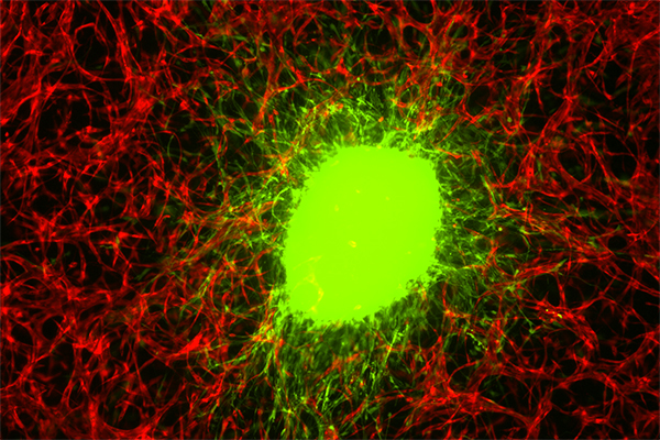
In order to study GBM more directly, Dai and his team 3D bioprinted an in vitro GBM tumor model with microenvironments that mimic the invasive tumor behaviors. As such, the model can act as brain tissue for the tumor cells to infiltrate. Explaining how the model was bioprinted, Dai states: “We use human brain blood vessel cells, and connect them with all the neurons, pericytes, astrocytes, the major cell types in the human brain.”Hydrogel was used as a matrix to hold these cells in place. “Then we use 3D printing to stack them in three dimensional fashion.”
The researchers sourced glioblastoma tumor stem cells from brain tumor patients with the help of Jenny Zou, a neurosurgeon, and Roland Friedel, a neuroscientist, at Mount Sinai’s medical school. These cells were then placed in the middle of the 3D bioprinted model, which is a few millimeters thick. Doing so allowed the researchers to observe how the brain tumor cells aggressively invade as they would do in patients, while also enabling long-term culture of the cells: “The 3D constructed model allows replicating 3D environment for the physiological tumor invasion and the antitreatment responses. The incorporation of a tumor spheroid within 3D matrix not only allows the GBM invasion but also increases the life span of the tumor spheroid. Long-term culture is required for tumor stem cell differentiation and therapeutic resistance development,” explained the researchers in the paper.
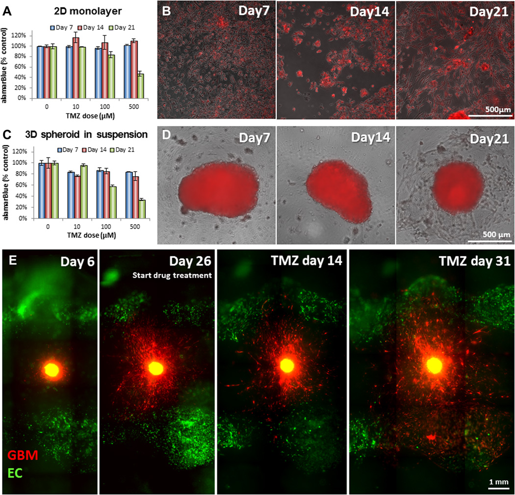
To accurately observe what was happening inside the 3D model without disrupting it, Xavier Intes, a biomedical engineer at Rensselaer Polytechnic Institute, scanned the sample using a laser, creating a 3D snapshot of the cellular structure. Combining this imaging technique with 3D bioprinting has allowed the researchers to assess how the tumor responded to a commonly used chemotherapy drug, temozolomide, evaluating its effectiveness in the process. They found that the drug did not work in the long term, matching the experience of patients with glioblastoma. “We treated the tumor with the same kind of drug you give to a patient when they undergo chemotherapy,” added Dai. “We monitored this chemotherapy over two months. And what we found was the chemotherapy was not able to kill the tumor.”
Using the model allowed the researchers to speed up the process of determining the effectiveness of a drug for treating glioblastoma. They found that temozolomide was able to kill glioblastoma cells in two-dimensional models, but when put into the 3D bioprinted model, the tumor grew back. As such, the method has been effective in identifying unsuccessful drugs early, therefore ensuring that only the most promising drugs move to animal, and eventually human, trials.
3D printing and brain tumors
The use of 3D printed models has proved a useful tool for investigating and treating brain tumors. They allow researchers and medical practitioners to accurately observe tumors either to better understand the disease or for presurgical planning.
For example, earlier this year 3D LifePrints, a UK-based medical technology company, 3D printed a tumor model to aid surgeons in the removal of a cancerous mass in six-year-old Leah Bennett. The model helped the surgeons with presurgical planning, thus increasing the chances of safe and effective removal.
In 2019 South Korean scientists also detailed how they used 3D bioprinting to create an ex vivo model emulating the characteristics of GBM tumors using extracted cells from human patients.
The research paper “High-resolution tomographic analysis of in vitro 3D glioblastoma tumor model under long-term drug treatment” is published in Science Advances. It is written by Mehmet S. Ozturk, Vivian K. Lee, Hongyan Zou, Roland H. Friedel, Xavier Intes, and Guohao Dai.
The nominations for the 2020 3D Printing Industry Awards are now open. Who do you think should make the shortlists for this year’s show? Have your say now.
Subscribe to the 3D Printing Industry newsletter for the latest news in additive manufacturing. You can also stay connected by following us on Twitter and liking us on Facebook.
Looking for a career in additive manufacturing? Visit 3D Printing Jobs for a selection of roles in the industry.
Featured image shows glioblastoma stem cells aggressively invade a model made of human brain cells and biomaterials. Photo via Guohao Dai.