The openVertebrate (oVert) project is scanning between 20,000 and 30,000 fluid-preserved vertebrate specimens from institutions across the US and making them freely available to 3D print on the MorphoSource platform, a platform hosted by Duke University.
Funded by a US National Science Foundation (NSF) grant, oVert has brought together research institutions who will CT scan the specimens, which constitute around 80% of living vertebrate genera, using a network of digitization centers across the US. Educators, scientists and designers will then be able to generate highly accurate models for teaching research or creation.
3D scanning, 3D printing and the oVert project
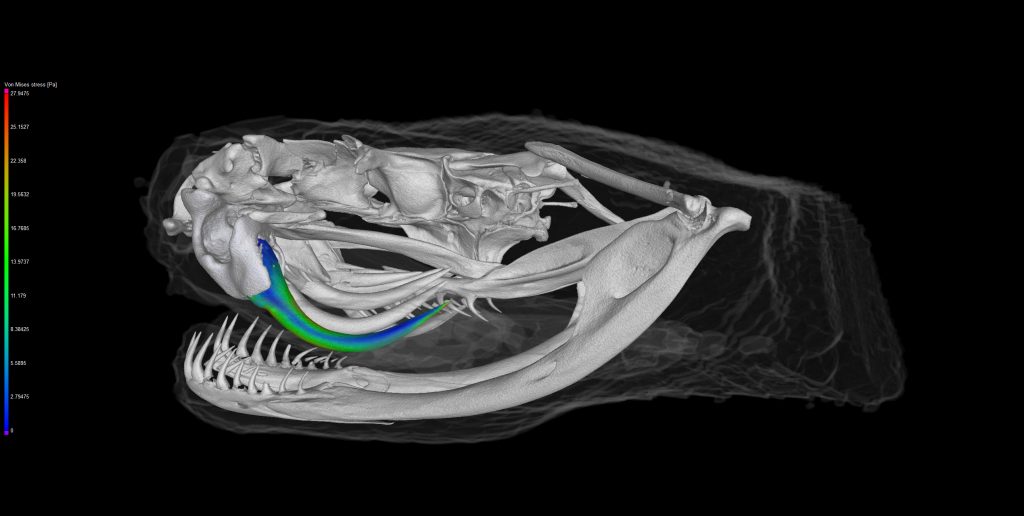
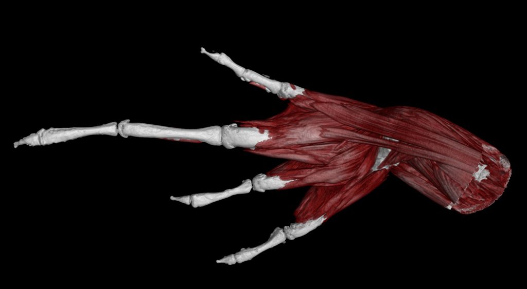
Scanning for the project, which began in August at the Florida Museum of Natural History, is being undertaken using x-ray computed tomography (CT) technology. This generates high-resolution digital anatomical data, which can then be represented as both 2D image stacks of internal anatomy and 3D surfaces of entire animals or individual body parts in a .ply format.
Through MorphoSource, oVert will provide both raw anatomical tomography data for specific research and a library of online 3D models which are ready to view, download, manipulate and 3D print primarily for research, education and other non-commercial use (in a similar manner to how Scan the World provides a 3D library of sculptures).
To accomplish this, oVert will work with Duke to upgrade the interface and functionality of MorphoSource by improving its capacity to explore media and integrate some metadata with iDigBio.
“CT scanning has been around for quite some time, and different researchers have been investing in CT for smaller individual projects…but the resulting stacked CT scans have never been freely available on this scale before,” said Luke Welton, a researcher responsible for the University of Kansas’ oVert project, which received $88,000 of NSF funding.
“You could model, say, the evolution of horses based on their teeth. Or model something rare, like a monitor lizard such as the little-known Varanus bitatawa or a Sphenodon punctatus…”

oVert in education, research and outreach
Current higher level research with oVert includes the study of patterns of relationships among both living and extinct vertebrates, testing hypotheses of morphological evolution, generating “structure-function” models for testing hypotheses, investigation of feeding and locomotion, and analysis of brain and nervous system anatomy.
“Having the digital data or a 3D replica there, it can really be a game changer for students who do better in a hands-on learning environment,” said Welton. He noted also that it was preferable to “use a 3D replica than actual specimens in certain settings to avoid wear and tear or have elements falling off.”
However, oVert is placing emphasis on education and training at all levels, from schools through to museum staff and researchers. In an effort to boost the use of digital specimens by STEM educators, oVert members are to teach high school and undergraduate students to create 3D anatomical models and will hold on-site workshops to help teachers incorporate 3D technology into lessons.
Led by David Blackburn and his team at the University of Florida, the oVert project also includes researchers at the Academy of Natural Sciences of Drexel University, California Academy of Sciences, Cornell University, Field Museum of Natural History, Harvard University, Louisiana State University, University of Kansas, the Scripps Institute of Oceanography, Texas A&M University, University of California Berkeley, University of Michigan, University of Texas Austin, University of Washington, Virginia Institute of Marine Science and Yale University.
Nominations for the second annual 3D Printing Industry Awards are now open. Make your selections now.
For more information on 3D object databases, subscribe to our free 3D Printing Industry newsletter, follow us on Twitter, and like us on Facebook.
Featured image shows oVert’s CT scan highlighting the heavy armor on the skull of this giant girdled lizard, Smaug giganteus. Florida Museum of Natural History image by Ed Stanley.