A paper from the Indian Institute of Technology (IIT) Delhi has demonstrated a 3D printable ink suitable for supporting the growth of cartilage. In a paradigm shift for the technology, the silk-based material differentiates between two different types of cartilage, potentially providing more specific treatment of joint damage.
3D bioprinting a replica for bone
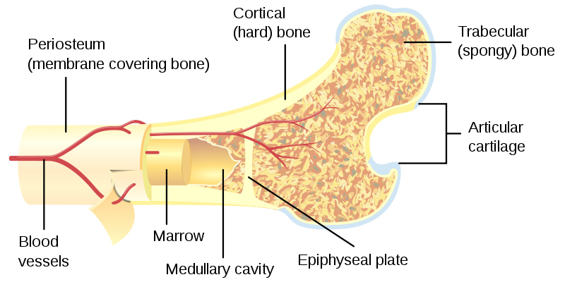
One of the key challenges in regenerative medicine, i.e. treatments using a patient’s own cells to heal damage, is an ability to replicate the complexity of tissue found in the body. When working with bone for example, there are at least two bone densities to contend with: the cortical, hard, outer shell, and the cancellous “spongy” tissue inside.
Cancellous bone tissue is made from a type of cartilage known as transient cartilage, that grows to become the long, interconnected cells in the center of a bone. Articular cartilage on the other hand is essential to cushioning the joints, allowing movement and protection from wear to the bone.
In pointing out that “cartilage has never been evaluated with an aim to distinguish between transient and articular”, the ITT study proposes a paradigm shift through six different conditions for cartilage production. In one part, a 3D bioprinted silk ink is used to support this growth, and shows a number of advantages over other types.
A “prospective tissue equivalent”
The 3D printable bioink is created by mixing silk fibroin protein with gelatin – a combination that ensures the ink becomes semi-solid when required. The mixture is sterilized in alcohol, 10X media cell culture solution, and 10% fetal bovine serum. It is then incubated at 37 °C for 15 min to ensure all ingredients dissolve uniformly.
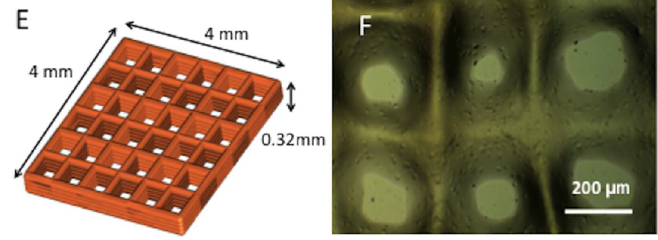
After this, the ink is immediately laden with bird-derived the bone marrow cells, and 3D printed into a grid design to replicate the natural cell structure in the body.
Two constructs are created – one that cultures into transient bone after day 4 in incubation; and another, more heavily laden with cells, that is still articular and viable for use up to day 14.
By comparing the proliferation and growth of cells in the variety of samples, researchers find that the 3D bioprinted ink shows similar qualities to those exhibited in human bone-cell development. Conclusions state that the material is “a prospective tissue equivalent” capable of activating organ growth in a “patient-specific manner”.
The full text of Elucidating role of silk-gelatin bioink to recapitulate articular cartilage differentiation in 3D bioprinted constructs is published online in Bioprinting journal, May 2017. It is co-authored by Shikha Chawla, Aditi Kumar, Prasad Admane, Amitabha Bandyopadhyay and Sourabh Ghosha.
The secret of silk
As a strong, biocompatible and degradable material, silk has found many uses in medicine to help heal the body. Previous silk related research reported by 3D Printing Industry includes its application in an ink to fix bone fractures, and to make the pins and screws used when fixing plates to broken bones.
Other cartilage bioprinting research includes Duke University’s creation of a meniscus to create life-like knee implants, and a paper from Chalmers University of Technology in Sweden for the treatment of osteoarthritis.
For more of the latest 3D printing related research and other news sign up to the 3D Printing Industry newsletter, like us on Facebook and follow us on Twitter.
Featured image: X-ray of cartilage damage in the knee. Image via medicalnewstoday
