3D bioprinting is gradually proving its efficacy to create functional, replacement tissues for the body. At present though it has only be proven in small animal models. If the technology is ever to reach clinical trials in human patients it will first need to be scaled up.
In Japan a collaboration between Keio University, Saga University, Kyushu Medical Center, Saga Hospital, Kyoto Prefectural University of Medicine and Cyfuse Biomedical K. K. has taken the next step toward this goal.
A recent study published by the associates in May 2019 demonstrates that 3D bioprinted vascular grafts can survive for long periods when implanted in pigs. The research has been described by the team as “an essential step towards a comprehensive and clinically relevant evaluation of human cell regeneration strategies at the pre-clinical stage.”
The Kenzan method of 3D bioprinting
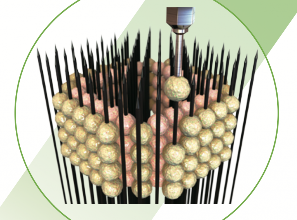
The 3D bioprinting method employed by the Keio-Saga team to make vascular grafts is called the Kenzan method. Commercialized exclusively by Cyfuse Biomedical K. K., in this technique spherical cell conglomerations known as spheroids are speared by a “Kenzan” array of needles. Left to culture the arrangement of these spheroids, e.g. a circular vessel/tube, merge together. At a certain stage they are removed from the needles then left to heal and close the remaining holes. What is left is a complete sample of tissue.
Over the years, the method has been used variously to create vascular implants trialed in rats, a mini liver model and, most recently, regenerative esophageal structures.
A first for maintaining patency
This latest paper titled “Development of an immunodeficient pig model allowing long-term accommodation of artificial human vascular tubes” increases the scale of Kenzan vascular trials conducted in rats, instead applied to so-called “operational immunodeficient pigs” or OIDPs.
In experimentation, the researchers undertook a procedure to suppress the reaction of the OIDP immune system in strict accordance with the recommendations in the Guide for the Care and Use of Laboratory Animals of the National Institutes of Health. Human stem-cell derived tubular tissues produced on a Cyfuse Biomedical 3D bioprinter were then implanted into the pigs, and studied by the team over a period of 3 months.
In two out of the six pigs in this trial, the patency of the artificial tissue was maintained for the duration of the experiment. At this stage, smooth muscle cells had started to form at at the implant, denoting the next stage of cell regeneration.
Though not all pigs shared the same results the study does show a high degree of success for the method. The researchers write, “It should be noted that this is the first report in which tissue-engineered grafts derived from human cells remained patent for > 1 month after xenotransplantation in a large animal model.”
The study concludes, “The birth of OIDPs without the rejection of human cell-derived organs symbolizes an essential step toward the comprehensive evaluation of preclinical cell regeneration strategies and contributes to the development of regenerative medicine using human organs, which is expected in the near future.”
Full results can be accessed online in nature communications journal. The study is co-authored by Manabu Itoh, Yosuke Mukae, Takahiro Kitsuka, Kenichi Arai, Anna Nakamura, Kazuyoshi Uchihashi, Shuji Toda, Kumika Matsubayashi, Jun-ichi Oyama, Koichi Node, Daisuke Kami, Satoshi Gojo, Shigeki Morita, Takahiro Nishida, Koichi Nakayama and Eiji Kobayashi.
Vote for your Research Team of the Year in the 2019 3D Printing Industry Awards.
For more of the latest research news subscribe to the 3D Printing Industry newsletter, follow us on Twitter and like us on Facebook. Seeking jobs in academia? Make your profile on 3D Printing Jobs, or advertise to find experts in your area.
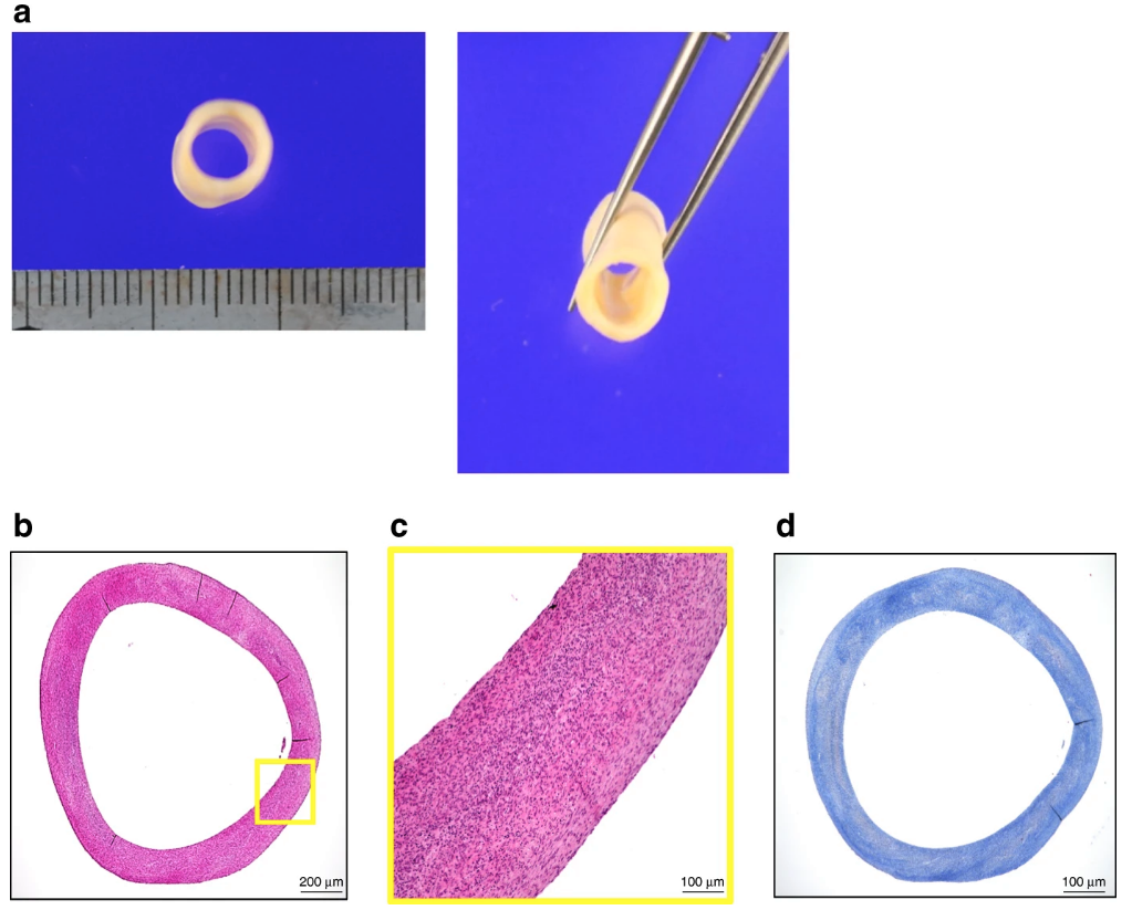
Featured image shows macroscopic photographs of the “human original 3D bioprinted tube” (HOBPT) used in this study. Image via nature communications
