3D printing is helping researchers in the fight against cancer. Around the world, experiments are applying the technology to help plan fail-safe tumor removal, and even to investigate new modes of treatment, such as targeted drug delivery.
At the Tomsk Cancer Research Institute of Tomsk Polytechnic University, Russia, a team has developed a new, 3D printable material, that helps to asses radiotherapy treatments.
“There is no need to explain that radiotherapy is a serious medical manipulation associated with certain risks,” comments Yuri Cherepennikov, a senior lecturer from Tomsk’s Division of Nuclear Fuel Cycles.
“The more carefully [a] treatment plan is elaborated and verified, the more efficient it will be, the less healthy tissues will be affected.”

3D printed phantom bodies
The 3D printed models made at Tomsk Polytechnic are called dosimetry phantoms. They are replica body parts used to asses which tissues will receive radiation. Cherepennikov explains, “We have developed a polymer material which is identical in density to the tissues of the body, and various additives allow creating analogues of a variety of tissues: bone, muscle, fat, and others.”
Through a combination of various materials the researchers are able to closely mimic the texture and density of an infected part of a patient’s body. This 3D printed replica is then given a required dosage of radiotherapy.
By assessing the outcome of the experiment, doctors can minimise the amount of damage done to a patient’s healthy tissue, and focus dosage onto the tumor.

A personal approach
Standard human models are already in use to assess radiation dosimetry, but the Tomk team is proposing a more personal approach. Cherepennikov adds, “…everyone understands that, for example, bones and muscles have different densities and interact differently with radiation.”
However, “We propose to create patient-specific models of the single body parts based on imaging data which are collected for each patient before radiotherapy, no additional manipulations are needed.”
Currently, the fabrication process of these patient-specific models can take up to two days in a laboratory, depending on the complexity of the phantom. But, through development, the team expect to be able to reduce personalized production down to just 10 hours.
Evidence of the lab’s current research into 3D printed dosimetry phantoms can be found online here.
For more of the latest 3D printing powered medical discoveries subscribe to the most widely read newsletter in the industry here, like 3D Printing Industry on Facebook and follow us on Twitter.
What medical research is leading the industry? Make your nominations for the 2018 3D Printing Industry Awards now.
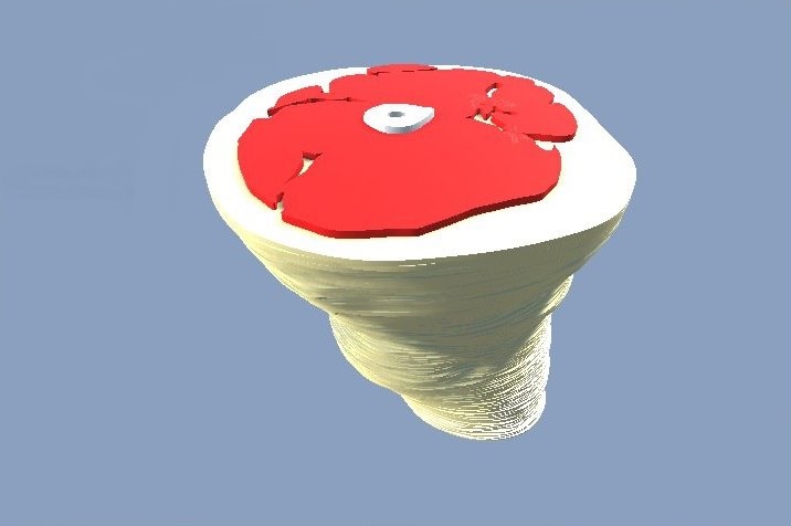
Featured image shows a false coloured image of cancer cells dividing. Image by McDougal Littell from Biology, 2008


