3D printing has a plethora of uses when it comes to body modification and functionality. We have seen a variety of assistive prosthetic devices, medical tools, and perhaps most excitingly, engineering geared toward exploring and repairing the inner workings of our bodies. One group of Japanese-based researchers have utilized bioprinting to create scaffold-free tubular tissues made of multicellular spheroids (MCS), which are composed of approximately 40% human umbilical vein endothelial cells, 10% human aortic smooth muscle cells, and 50% normal human dermal fibroblasts. The team produced 500 robotically configured and 3D printed MCS-based tubular tissues around the structure of a needle, which acts as the necessary scaffold support needed in the early stages of creation.
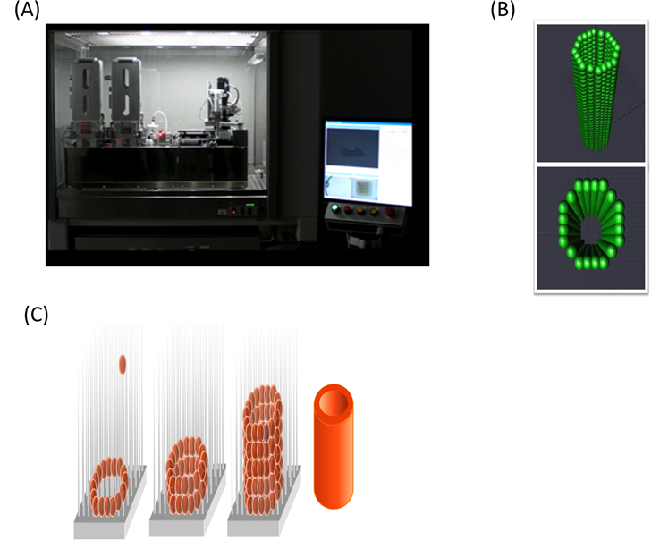
 Before being able to accurately produce this tissue material, there is meticulous preliminary 3D design work that must be done. The team must first check the spheroids’ sizes, shapes, and circle rates via design software, making sure that the physical properties of the MCS-based tissue structure match those found in the body. After the design is perfected, the spheroids are handled gently, picked up and loaded into to the needle-array. After four days of curing on the scaffolding needle, the needle-array is then removed, since the fusion between it and the MCSs has now bonded to create a structurally sound and scaffold-free tubular tissue material. The entire process can be observed in their project video (shown below). The 3D printed design seemed promising, but the next step for these scientists was to find a non-human animal with similar cells to human beings, which is where they became dependent on the classic laboratory pet, the rat.
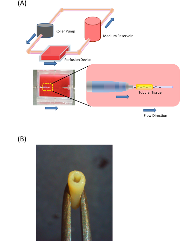
 The outcome of this MCS-based method was quite successful, as the team was able to create a tubular structure without any scaffolding support, and have already tested the tissue implant in the abdominal aortas of their laboratory rats. Before being implanted in the rats, the bioprinted tubular tissue is cultured in a perfusion system. After the initial experimentation with the Japanese nude rats (chosen, of course, because their cells are similar to those of human origin), the team was able notice that the cells helped produce an enlarged lumen area and a size decrease in the wall area. These results led to an on-going remodeling of the MCSs, where further experimentation will be performed to elucidate the mechanism of endothelialization in both short-term and long-term scenarios.
The complete research, titled “Scaffold-Free Tubular Tissues Created by a Bio-3D Printer Undergo Remodeling and Endothelialization when Implanted in Rat Aortae”, can be read in the journal PLoS ONE. While the process is still underway, this team of bright bio-engineers are taking us one stop closer to bioprinting MCS-based tissue that will hopefully make its way over to aid the internal structure of the human body.