Scientists from the University of Leuven (KU Leuven), and VIB, a life sciences research institute based in Belgium, have developed a multi-color optical method for 3D imaging surfaces and structures.
The research paper published in the BMC explains, “Current mesoscale 3D imaging techniques are limited to transparent or cleared samples or require the use of X-rays. This is a severe limitation for many research areas, as the 3D color surface morphology of opaque samples cannot be assessed.”
Dubbed as “ALMOST” (A Label-free Multicolor Optical Surface Tomography), this method can be used for digital repositories of zoological and botanical collections which can then be 3D printed.
Medical and zoological 3D imaging
Micro and mesoscale 3D imaging have allowed for the analysis of microorganisms, embryos, and organs within biomedical research.
Nevertheless, according to Sebastian Munck, a light microscopy expert from VIB-KU Leuven, 3D imaging requires making a sample transparent using chemical ‘clearing’ methods. Such methods are “time intensive and can’t be applied to every type of sample.”
“Moreover, if you want to study surface morphology or color, optically clearing is counterproductive.”
Thus, in order to 3D image morphological details of non-transparent samples, the researchers combined optical projection tomography (OPT) and oblique illumination – a technique whereby light is projected at an angle to reveal features with higher contrast. Furthermore, color filters and a projection algorithm were used together with 3D rendering software to eliminate the need for clearing methods.
“ALMOST opens the possibility for longitudinal imaging of unaltered, live samples,” the study states.
“It reveals complementary information to transmission and fluorescence, it poses an ideal supplementary approach to well-established OPT and light sheet modalities and allows imaging of the sample color, which is lost in X-ray-based techniques like micro-CT.”
The ALMOST method
To validate their method, the VIB-KU Leuven team reconstructed the 3D color surface from a diverse set of samples including an electrical resistor, seed cones of the dawn redwood, Lego figurines (which we compare with micro-CT), and a shell of a sea snail.
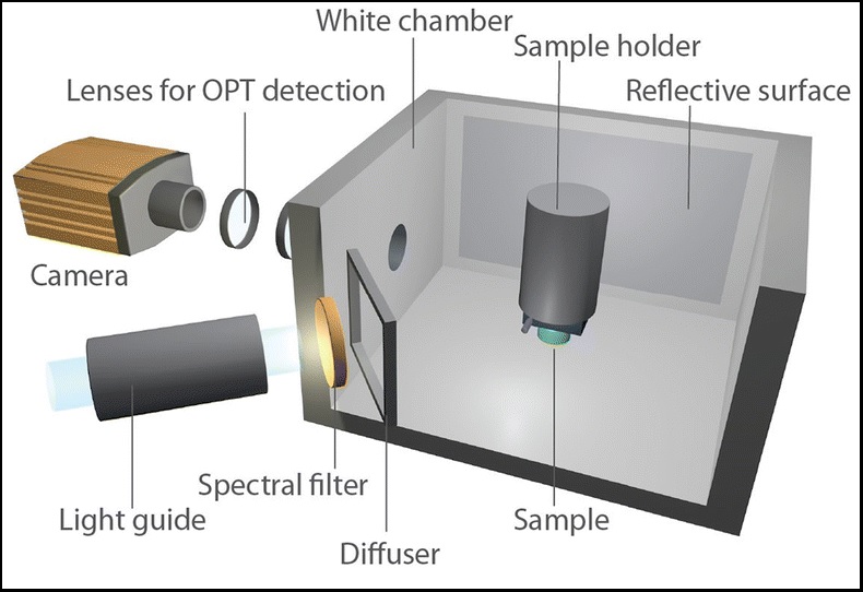
Using a SkyScan 3001 M OPT scanner, manufactured at Bruker, images of the object were captured within the illumination chamber. This chamber was built with white paper and aluminum foil, an unfocused light source of LED goosenecks, and a diffuser made of milk glass.
The imaging chamber was designed to obtain images of the sample as if it were a self-radiant object. The study adds, “this is important, as this allows the images [to] mimic an absorbing or fluorescent sample and can thus be analyzed in a similar way.”
The researchers concluded, “to render shapes in 3D acquired with reflective light imaging, it is essential that the illumination system provides a difference between the background intensity of light and the reflected light that has interacted with the sample.”
“In addition, for 3D visualization, the background will typically be rendered transparent to reveal the 3D shape of the sample by a process called ray tracing. Our approach of using a white background achieves this for any non-white (or less bright) samples.”
“A Label-free Multicolor Optical Surface Tomography (ALMOST) imaging method for nontransparent 3D samples,” is co-authored by Axelle Kerstens, Nikky Corthout, Benjamin Pavie, Zengjin Huang, Frank Vernaillen, Greetje Vande Velde and Sebastian Munck.
Who has made the most significant academic contribution to additive manufacturing?Nominate for the upcoming 3D Printing Industry Awards 2019.
For the latest 3D printing research, subscribe to the 3D Printing Industry newsletter, follow us on Twitter and like us on Facebook.
Looking for a fresh start this year? Visit 3D Printing Jobs to commence your career in additive manufacturing.
Featured image shows a shell in 3D color created from ALMOST. Clip via VIB-KU Leuven.