One major way that the medical industry has been impacted by 3D printing technology is with patient-specific 3D modeling, which has allowed doctors to accurately prepare for intensive procedures. BMC Anesthesiology, an open access, peer-reviewed journal that focuses on all aspects of anesthesiology, published a case report from the Beijing, China-based Peking University Third Hospital’s Department of Anesthesiology on January 19, 2016, entitled “Three-dimensional printing as an aid to airway evaluation after tracheotomy in a patient with laryngeal carcinoma.” The 77-year-old patient involved in the study had undergone a total laryngectomy, which is the full removal of the larynx and separation of the airway from the mouth, nose and esophagus, an extremely difficult and dangerous procedure.
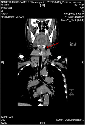
An evaluation had shown a fistula forming at the place of the tracheotomy incision. Using at CT scanner and Materialise Mimics software, which is used for the segmentation of 3D medical images, the research team obtained a 3D model of the patients trachea. After exporting the 3D reconstructed image as an STL file, the team used a MakerBot Replicator 2 to 3D print patient-specific inner and outer tracheal diameters. The models were printed in white PLA and were moderately hard, which met the needs of the researchers.
Using the various tracheal 3D models, the research team, which included Bin Han, Yajie Liu, Xiaoqing Zhang, and Jun Wang, found a few glaring issues that need immediate attention. They found slight scar retraction around the stoma that was created by the tracheostomy, and also slight stenosis below the stoma in the trachea too. Using the inner tracheal model, the team created an anesthesia plan for the patient, and then practiced the incubation steps to prepare for the procedure.
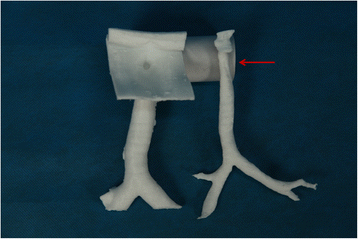
According to the study, there were some drawbacks to the 3D printing technique, as it could not tackle the need for color variations in the tracheal mucosa, which help distinguish inflammation of the airway wall. Still, the team had an extremely positive experience with 3D printing technology.

3D printing was a groundbreaking technique and has matured. The successful outcomes of our case suggest the great practicability of preanesthesia planning, and more widespread use is encouraged,” they write in their study. “In the future, this technology could be better used not only to evaluate the difficult airway but also create artificial tracheal tubes for specific patients, thereby realizing individualized anesthesia.”