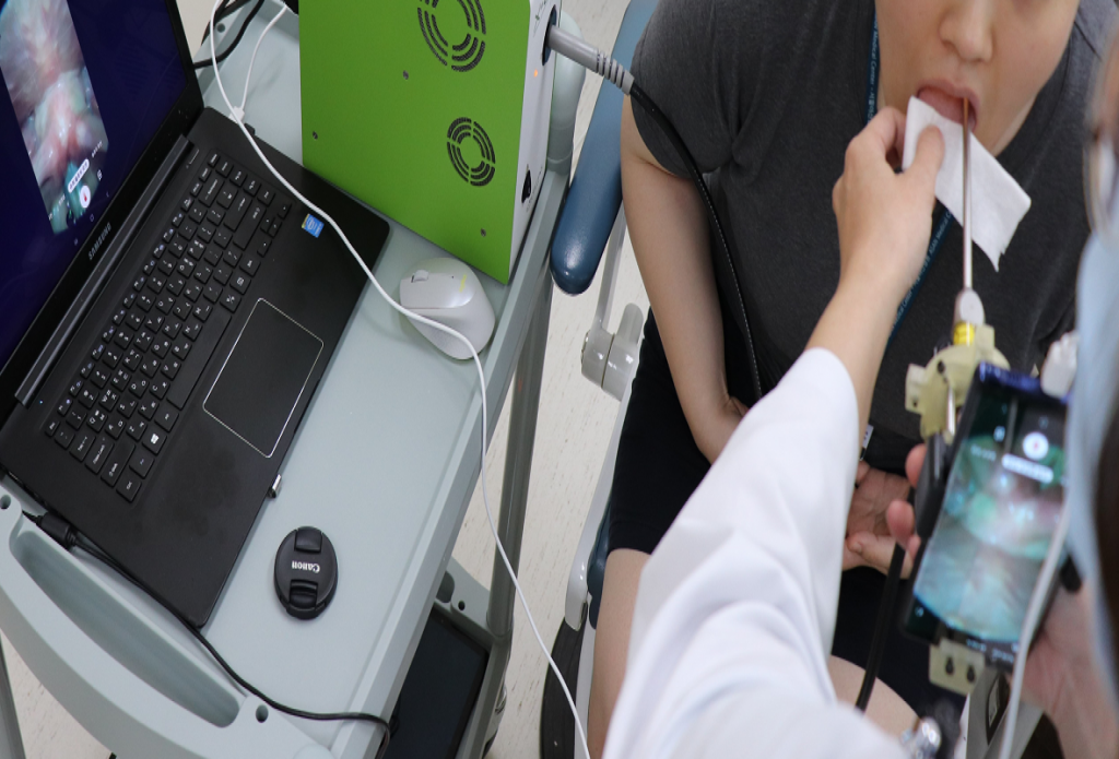
A group of Korean scientists have developed a low-cost 3D printed adapter that turns everyday smartphones into endoscopic vocal cord disease diagnostic tools.
Once fitted with a laryngoscope, the team’s SLM 3D printed device is capable of capturing and relaying high-quality images of a patient’s throat, using any modern phone’s built-in camera. Given that the mobile add-on costs much less than conventional digital imaging systems, it could prove to be an accessible point-of-care analysis tool in future, for those clinicians operating in developing countries.
“This simple, low-cost adaptor promises to reduce the cost of clinical imaging by an order of magnitude when it can be used as a substitute for standard high-speed digital imaging (HSDI),” said the team in their paper. “The device demonstrates the promise and emerging capabilities of clinical diagnostic tools that incorporate commodity smartphone technology to bring point-of-care diagnosis to remote locations.”
Endoscopies for the people
For devices that are essentially long, thin, flexible tubes, endoscopes are a vital tool in the armory of medics, as they enable the visualization of otherwise-obscured internal organs. In areas like throat disease, such devices not only allow doctors to perform minimally-invasive surgeries, but to achieve more specific diagnoses via clinical voice evaluation.
Through vocal cord assessments, clinicians essentially aim to detect irregularities which correlate with disorders, but given that most voices operate at 80-240 Hz, using standard 60fps video makes this difficult. While improved HSDI devices have already been developed, they’re often more expensive, require specialized knowledge to use and have large data footprints that need post-processing.
As a result, endoscopic diagnosis and training is largely inaccessible to patients and staff in developing countries, and this can lead to poorly-performed or even fatal procedures, thus the creation of a more affordable and manageable HSDI imaging system has become an urgent issue for those in the third world seeking an assessment of their ailments.
As many smartphones now possess the processing power of small computers, they have shown considerable promise in similar areas before, with researchers using them to conduct everything from biological analysis to blood testing, and taking inspiration from these approaches, the Korean team have opted to develop a novel low-cost endoscopic mobile adaptor.
A higher-res oral procedure?
Designed for use with a standard laryngoscope, the researchers’ device consists of a holder, lens tube, lenses and endoscopic mount. To optimize the performance of their smartphone accessory, the team created its holders and connectors with a Stratasys Objet260 3D printer, and by holding its lens system in place, the part allows it to magnify the images captured by its eyepiece and camera respectively.
Additionally, rather than using achromatic lenses for image magnification, the device was fitted with two low-cost 50 mm and 15 mm biconvex lenses, providing it with 1.5x zoom capabilities. Once ready, the scientists attached their mountable endoscope to a Samsung Galaxy Note 9 smartphone, and got an otolaryngologist to test it for them on four consenting patients at the Korean Asan Medical Center.
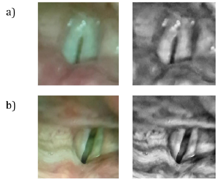
At peak performance, the system proved capable of operating at a resolution of 43.478 μm from a distance of 15 mm, but accuracy demonstrably decreased as the device was moved further away. Compared to commercial $10,000 HSDI systems, the team’s adapter also captured a much lower level of detail, recording images at 940 fps rather than the 4000 to 8000 fps seen in more standard devices.
Although the researchers concluded that the resolution and frame rate drawbacks of their adaptor currently make it insufficient for capturing the detail necessary for diagnosing many vocal cord conditions, they suggest that optical shielding and AI–based image processing could be used to overcome its performance and acquisition-related issues in future.
“Our results demonstrate that it is possible to process clinical images for useful diagnostic reference values of healthy subjects,” concluded the researchers in their paper. “In future, the development of more compact and optically-shielded adaptors will be required… to improve the distribution of healthcare capabilities.”
3D printing in throat diagnosis
Over the last twelve months, 3D printing has found significant applications in the diagnosis and treatment of throat-related illnesses. Researchers at ETH Zurich have developed additive manufactured bioresorbable stents, which are designed to provide a less invasive means of oxygen relief to those suffering from asphyxiation.
Scientists at the Pohang University of Science and Technology (POSTECH), meanwhile, have 3D printed novel stents that help ease the symptoms of esophagitis in radiotherapy patients. Based on a throat cell-derived bio-ink, the team’s inflammation-reducing devices could also be loaded with other cells in future, as a means of treating local injuries.
In other more experimental applications, a team at the University of Oregon (UoM) has 3D printed a microscopic device that enables control over the impulses that drive vocalizations in songbirds. By modulating electrical signals sent to the brain or spinal cords of their zebra finch test subjects, the team were able to evoke distinct predetermined birdsongs.
The researchers’ findings are detailed in their paper titled “A Portable Smartphone-Based Laryngoscope System for High-Speed Vocal Cord Imaging of Patients With Throat Disorders: Instrument Validation Study.” The study was co-authored by Youngkyu Kim, Jeongmin Oh, Seung-Ho Choi, Ahra Jung, June-Goo Lee, Yoon Se Lee and Jun Ki Kim.
During the project, research was conducted collaboratively between the Eulji University School of Medicine, University of Ulsan and Asan Medical Center.
To stay up to date with the latest 3D printing news, don’t forget to subscribe to the 3D Printing Industry newsletter or follow us on Twitter or liking our page on Facebook.
For a deeper-dive into additive manufacturing, you can now subscribe to our Youtube channel, featuring discussion, de-briefs and shots of 3D printing in-action.
Featured image shows a diagram of the Korean team’s 3D printed endoscopic smartphone accessory. Image via the JMIR Mhealth Uhealth journal.
