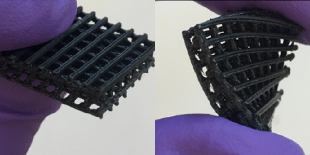
New 3D bio-printing research shows that combining materials could be the secret to 3D printing bones and organs. A paper from Case Western Reserve University (CWRU) in Cleveland, Ohio, describes research into the strength and bio-compatibility of a 3D printable material made from a mix of TPU, PLA, and graphene oxide.

TPU & PLA: the basic ingredients
TPU (thermoplastic polyurethane) is a material that is particularly useful in biomedical research. It is flexible, transparent, and compatible with living cells. By combining TPU with PLA, a material available in a bio-compatible form and one of most commonly used material in 3D printing, researchers can produce objects that have strength as well as flexibility.

This blend of qualities is particularly useful when creating something for the body, as tissue and bone have complex structures that can’t easily be replicated by a single synthetic material. Taking a bone for example – the ball joint sockets are by nature much stronger, and the middle of a bone is relatively flexible to allow for some strain.
In some ways TPU/PLA is a kind of base ingredient for biomedical material research, and adding other substances to the mix can not only enhance and modify its qualities, but also make the material even more ‘friendly’ to cells.
Enter graphene oxide
As a two-dimensional form of carbon (only a single atom in thickness), graphene is probably best known for its theoretically high strength. By oxidising the material, it then gains some oxygenated qualities, such as an ability to disperse in water. As it’s no secret that the body is made up almost entirely of water, it then becomes clear why instilling this hydrophilic feature into a 3D printed scaffold would be beneficial to biomedical research.
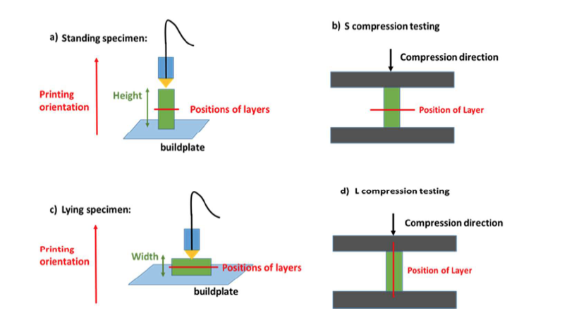
The primary reason for using graphene oxide (GO) alongside TPU/PLA in CWRU’s research is however to improve the mechanical properties of a material, so it performs better under pressure along the length and width of an object.

Research findings
CWRU’s conclusions show that adding a controlled amount of GO to TPU/PLA is not toxic to living cells, and ultimately improves the rate at which the cells grow and spread. The finished combination is also 167% stronger than TPU/PLA alone when compressed, and 75.5% better when stretched.
3DPI asked another expert for their professional opinion on the research. Dr. Guillaume Poize, biomedical research director at iMakr, explains that the enhanced properties of TPU/PLA/GO are of particular interest as ‘you can print on a bigger scale, and also produce the same objects with less materials [and cost]’, both of which are necessary if 3D bio-printing is become a reliable and viable option for medicine and bio-engineering.
If you’d like to show your support for the team at CWRU you can nominate this research for the Academic category of the 2017 3D Printing Industry Awards here.
Featured image shows bamboo scaffolding used in building construction. Photo via: Jellybean on flickr


