A recent progress report published in the journal Advanced Healthcare Materials details the results of various experiments that have utilized the Kenzan method of 3D bioprinting to produce cell constructs for transplantation. The paper is intended to give readers an overview of the state of the art technology in 2020.
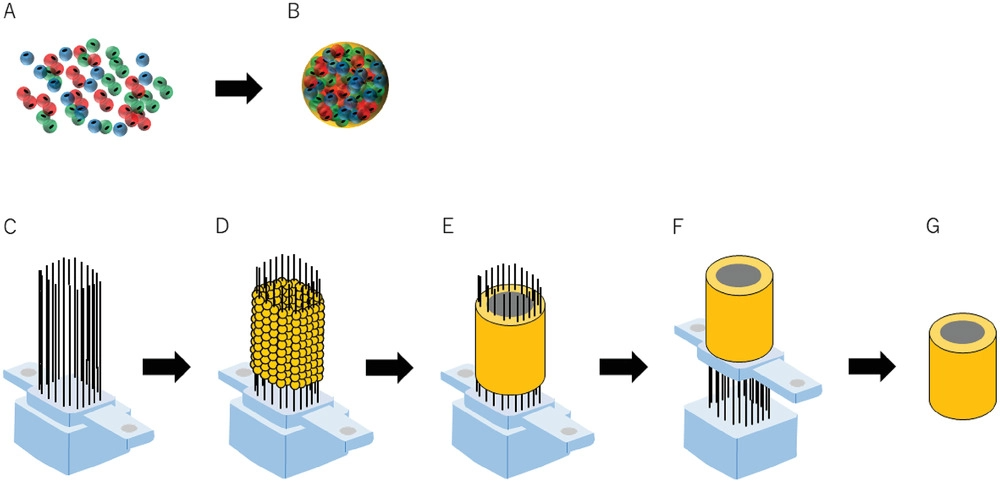
Since the turn of the millennium, research into 3D bioprinting has gained some serious traction. One of the more developed methods of bioprinting, the Kenzan method, involves skewering a culture of cells called spheroids on an array of microneedles (the Kenzan) and waiting for them to partially fuse. Once the cells are able to support their own structure the needles are retracted and the cells are nurtured and grown into a usable tissue patch.
According to the pioneers of the method at Saga University, it is the only method of 3D bioprinting surgically transplantable cell-only structures without the need for artificial additives. The cell-only nature of the Kenzan method qualifies it as a ‘scaffold-free’ process as the only supporting structure of the bioink is the array of microneedles.
Peripheral nerve reconstruction
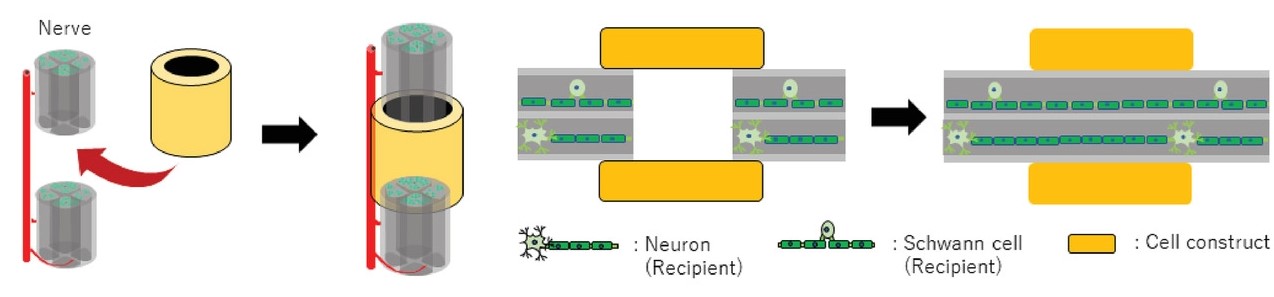
If the nerve gap lengthens significantly after a peripheral nerve injury, an autologous nerve transplant may be required. This first-line-of-defense treatment does have its downsides, however. The healing over of the transplant may result in inconsistencies and neuroma formation which is problematic and requires further treatment. Therefore, Yurie et al. attempted to reconstruct scaffold-free cellular nerve conduits for the peripheral nerve of a rat.
Using dermal fibroblasts – the spheroids in this case – taken exclusively from a human, the team 3D bioprinted the tubular cell constructs and implanted them at the sciatic nerve of a rat. The nerves were still connected eight weeks after the procedure and functional neurons could be observed in the nerve gap. The researchers concluded that the transplanted tubular structures had promoted functional nerve reconstruction at the site of the sciatic nerve.
Functional heart tissue reconstruction
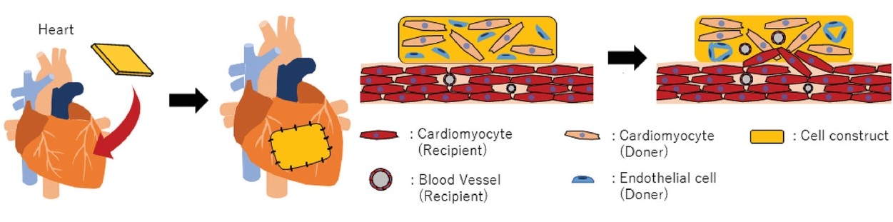
Today’s treatments for heart failure tend to only relieve symptoms for a relatively short time and delay the inevitable, as the reconstruction or healing of functional heart tissue is not yet established. Ong et al. attempted to use the Kenzan method to reconstruct functional, healthy cardiac tissue to mend a (broken) rat heart.
The spheroids in this experiment included cardiomyocytes derived from pluripotent stem cells and ventricular cardiac fibroblasts – both sourced from humans. Once transplanted, the researchers observed a spontaneous pulsation of the cell construct. Full engraftment was confirmed a week after the surgery and the transplanted heart tissue was found to contribute directly to the functionality of a rat heart.
Esophagus reconstruction
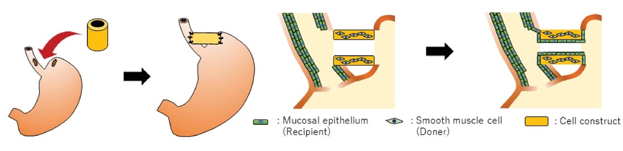
Current methods of esophageal reconstruction post-esophagectomy more-often-than-not result in complications and have a high mortality rate. To address this, Takeoka et al. attempted to 3D bioprint esophagus-like constructs to be transplanted into rats and observed the results.
The spheroids used in this experiment included human dermal fibroblasts and human esophageal smooth muscle cells. Once the tubular tissue structures were fabricated, they were implanted and fully engrafted after 30 days. The researchers found that the rats could successfully pass food through their new foodpipes into their stomachs without any complications. The team concluded that the experiment “exhibited promise” and expects to scale this research up to a human procedure one day.
A detailed explanation of the Kenzan method and its extensive applications can be found in the paper titled ‘Scaffold‐Free Bio‐3D Printing Using Spheroids as “Bio‐Inks” for Tissue (Re‐)Construction and Drug Response Tests’. It is co-authored by Daiki Murata, Kenichi Arai, and Koichi Nakayama.
While the Kenzan method is unique in its scaffold-free nature, other methods of 3D bioprinting have also seen success recently. In Korea, CLECELL, a 3D bioprinting startup, has developed a respiratory epithelium model using its proprietary 3D bioprinting technology. The team intends to use the model as a testbed for various viruses to better understand their behavior. Elsewhere, on the ISS, nScrypt has completed the first functional 3D bioprinting experiment in the microgravity of space – a human knee meniscus.
The nominations for the 2020 3D Printing Industry Awards are now open. Who do you think should make the shortlists for this year’s show? Have your say now.
Subscribe to the 3D Printing Industry newsletter for the latest news in additive manufacturing. You can also stay connected by following us on Twitter and liking us on Facebook.
Looking for a career in additive manufacturing? Visit 3D Printing Jobs for a selection of roles in the industry.
Featured image shows the Kenzan method and its many applications. Image via Saga University.